Astigmatism: signs, diagnosis, treatment
Astigmatism
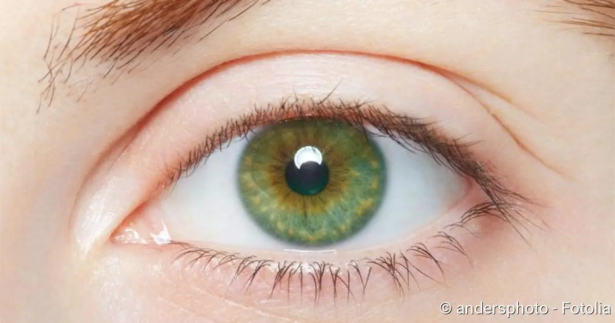
In astigmatism, an irregular shape of the cornea of the eye causes the incident light to be imaged distorted on the retina (Source). As a result, the patients affected with this condition have their vision blurred (Source). There are many causes of astigmatism (Sources), but it is often congenital (Source). It can often be corrected with special visual aids (Source) of different types (Source). Read more about astigmatism.
ICD codes for this condition are H18 H52
Description
The cornea is the foremost part of the eyeball that lies in front of the pupil. It is slightly oval in shape, slightly smaller than a small coin, and about half a millimeter thick (Source). Since it rests on the round eyeball, it is itself spherically curved, much like a contact lens.
What is astigmatism?
We speak of astigmatism (inaccurately: “corneal curvature”) when the cornea is not evenly curved. This anomaly is called “astigmatism”.
Normally, the cornea, together with the lens of the eye, ensures that the parallel incident rays of light are bundled and focused on a single point on the retina (focal point). This enables sharp vision.
In astigmatism, however, the cornea is unevenly curved, which means that the light cannot be properly focused. In some places, incoming light rays are bundled more strongly than in others. As a result, they do not merge into a single point on the retina: no single clear point is imaged on the retina – vision is blurred.
What types of astigmatism are there?
In regular astigmatism, incident light rays are imaged on vertical focal lines (“rod”). This form of astigmatism can be further subdivided, but it is mainly relevant for the optician to be able to produce a perfectly fitting visual aid.
Irregular is a curvature of the cornea whose optical planes are not perpendicular to each other. To put it more simply: While there is still a certain order in regular astigmatism, an irregular astigmatism sometimes has no recognizable system at all. In extreme forms, such as corneal scarring, the light is deflected in various directions so that there are hardly any focal lines left. The treatment of irregular astigmatism is therefore more difficult.
Astigmatism can also be judged by where the focal lines lie in relation to the retina. Often one is in the retinal plane, but the other is in front of it (astigmatism myopicus simplex) or behind it (astigmatism hyperopicus simplex). There can also be a focal line in front of it and the other one behind it (astigmatism mixtus). Sometimes there is farsightedness or nearsightedness (hyperopia or myopia) in addition to astigmatism: “Astigmatism compositus” is what the specialist calls this.
Corneal Curvature Symptoms
- blurred vision near and far. In contrast to this, with short-sightedness or farsightedness, only near vision or only far vision is impaired.
- Headache and eye pain
- in children, possibly a permanent decrease in vision
Many patients primarily complain of headaches and eye pain with slight astigmatism. In contrast, symptoms of impaired vision often appear later or not at all. This is because the eye is constantly trying to correct the blurred image by changing the shape of the lens, which in the long run strains certain eye muscles, eventually resulting in headaches and eye irritation.
When vision problems occur, the environment not only appears blurred to those affected, but usually also distorted. Because there is no focal point on the retina, but rather focal lines, they tend to see punctiform structures as stripes or rods. This also explains the term “astigmatism”.
If it is more pronounced, congenital astigmatism in children leads to a so-called weakness of vision (amblyopia) if left untreated. In medicine, this is the name given to a visual impairment that occurs because vision is not properly “learned”. Since a sharp image never falls on the retina, no correct sensory perception can be transmitted via the optic nerve to the brain, where the data is processed. If a congenital visual defect lasts too long, the brain adjusts to it and then makes normal vision impossible, even if the actual visual defect is eventually corrected.
Causes and risk factors
In many cases, astigmatism is congenital. Occasionally it is also hereditary – the astigmatism is then manifested in several family members. An example of congenital astigmatism is the so-called keratoglobus, in which the cornea is curved forward and thinned out.
Under certain circumstances, astigmatism may not occur until adulthood. Then it is caused by, for example:
- Ulcers and scars on the cornea (caused by injuries, inflammations and infections of the cornea)
- Corneal cone (keratoconus): Here the cornea curves in several thrusts to a cone, which is usually noticeable between the age of 20 and 30.
- surgical interventions on the eye, such as operations to treat glaucoma
Examinations and Diagnosis
The ophthalmologist can clarify suspected corneal astigmatism using various examination methods. The type and extent of astigmatism can also be determined.
For example, the visual defect can be determined by means of so-called objective refraction. This involves projecting an infrared image onto the patient’s ocular fundus and simultaneously measuring whether this image is sharp. If this is not the case, various lenses are placed in front of the eye until a sharp image is obtained. This allows the examiner to draw conclusions about the type of visual defect.
If it is clear that astigmatism is present, the cornea can be measured more precisely and astigmatism can thus be determined more precisely. This is done with an ophthalmometer, for example. The device, which remotely resembles a microscope, projects a hollow cross and a reticule onto the cornea of the affected person: On an ideally shaped cornea, both would lie exactly on top of each other. With astigmatism, on the other hand, the crosses shift against each other, and the more pronounced astigmatism, the more the crosses shift against each other. Values for the radius of curvature and the refractive power of the cornea can be determined very precisely in this way.
In the case of irregular astigmatism, however, this diagnostic device reaches its limits. Here, a computer-controlled corneal topographer (video keratoscopy) is used to analyze the refractive power of the entire corneal surface. This examination provides the most accurate data on the type and extent of astigmatism.
For those affected who want to get an impression of their own astigmatism, there are special graphics that make this possible. To do so, they have to look at a so-called astigmatism sun wheel or ray figures from different distances. In the case of astigmatism, typical distortions then result. However, such graphics are not a reliable astigmatism test.
If astigmatism has been specified by different devices, finally the subjective refraction follows: Here the cooperation of the patient is required. While he is looking at visual charts, the ophthalmologist holds various visual aids in front of his eyes one after the other. The patient must now say which visual aid he uses to see the visual charts most sharply. Once this has been clarified, nothing more stands in the way of treatment.
Treatment Of Astigmatism
If the angle and the refractive error of a corneal curvature are known, one can try to compensate for the visual defect with suitable visual aids:
- Spectacle lenses with cylindrical grinding (cylinder lenses)
- soft, appropriately curved contact lenses, which align themselves on the curved cornea
- hard contact lenses, which bend the cornea correctly
The first glance through spectacle lenses is both a blessing and a shock for most people with this condition. Although they can now see clearly, the world appears unusually curved. And the later the astigmatism is corrected, the slower the eye gets used to the visual aid. It is not uncommon for this adaptation to be accompanied by headaches.
Astigmatism: laser surgery
Besides visual aids, there is also the possibility of laser treatment of astigmatism. The hot light beam removes the irregularities in the cornea and thus creates an even surface. The ophthalmologist has to decide whether a laser procedure is suitable from case to case.
Astigmatism: lens replacement
Another surgical treatment approach is the correction of this condition with a new lens. The cornea is left as it is, instead the eye lens is removed and replaced by an artificial lens (intraocular lens). It is shaped to compensate for this condition as much as possible. This procedure is usually only used in cases of a very pronounced condition.
Corneal transplant
In rare cases, neither visual aids nor the surgical procedures mentioned above can help. The last resort is corneal transplantation. Here, the curved cornea is removed and an intact donor cornea is implanted as a replacement.
Prognosis And Course of Disease
Compared to myopia or hyperopia, this condition is a relatively complicated visual defect that is therefore not so easy to treat. Unfortunately, the existing visual aids do not offer a 100 percent satisfactory result either. Normally, it does not progress but remains constant. An exception is keratoconus: With this variant, this condition increases more and more.
Sources:
- Deetjen, P. & Speckmann E.-J., & Hescheler, J.: Physiology, Urban & Fischer Verlag/Elsevier GmbH, 4th edition 2004
- Lang, G.K. et al: Ophthalmology, Georg Thieme Verlag, 4th edition 2008
- Grehn, F.: Ophthalmology, Springer Verlag, 31st edition 2011