Atheroma: Causes, symptoms, treatment
Atheroma: Causes, symptoms, treatment
Atheroma: Description
Atheroma is what doctors call to a “bump” surrounded by a layer of skin, which is mainly filled with skin cells and fat. Such filled cavities, which develop due to an obstructed gland, are also called retention cysts – in this case it is a trichilemmal cyst (“hair root sheath cyst”). Colloquially, the atheroma is also called a grotto bag.
Atheromas develop in the area of the hair root, and therefore increasingly in areas of the body with many hairs. Up to 90 percent of them are found on the hairy scalp, but sometimes also on the neck, face, stomach or intimate area.
An atheroma usually becomes one to two centimetres in size, in individual cases even larger. The cysts can occur individually or in groups. In principle, any person can develop atheromas, but some have a hereditary predisposition – in which case many cysts are often found on the scalp simultaneously.
Occasionally the so-called epidermoid cyst is also called an atheroma. These pea to plum-sized nodes also develop from hair roots, but from their uppermost part (infundibulum). They contain mainly peeled horn material, which is piled on top of each other. In contrast, the “real” atheroma is mainly filled with a very greasy substance.
Atheroma: Symptoms
An atheroma is an externally visible elevation in the skin. It is full to bursting, but more elastic than hard. Some atheromas can be moved under the skin. After an inflammation, however, they can also sit firmly on the scalp – this is where 90 percent of atheromas are found. However, they can also occur in other parts of the body, such as the neck, chest, abdomen or genital area. If several atheromas are present at the same time, they sometimes lie together in groups.
Most atheromas have a diameter of one to two centimetres. However, they can swell up to the size of a chicken egg – in rare cases even to the size of a tennis ball. In larger cysts, the skin that spans them is stretched out to a great extent. This leads to the hairs growing here being further apart or missing altogether. In some cases, a grey or black dot can be seen on the surface of the atheroma.
Normally an atheroma has the colour of the surrounding skin. It stands out primarily because of its protrusion. As long as it has not become inflamed, it is painless and causes little discomfort. However, depending on the fit and size, those affected may find it cosmetically disturbing.
In case of inflammation, the skin in the area of the atheroma reddens, it swells and touching or light pressure causes pain. If additional pus collects inside the atheroma capsule, it is an abscess.
Atheroma: causes and risk factors
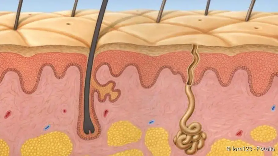
An atheroma usually develops in the scalp from a hair root (follicle) – more precisely from the narrow channel where the area of hair is located that is still hidden under the skin. With each hair a small sebaceous gland opens into this channel. It ensures that the hair is coated with a film of oily liquid – sebum. If the sebaceous glands are very active, the hair quickly becomes greasy.
The excretory duct of the sebaceous gland can be blocked in a certain area, the so-called isthmus, for example by small fat crystals or skin cells. The sebum can no longer flow off unhindered, but the gland continues to produce it. Gradually the sebum accumulates and the hair root is pumped up to a round “bubble” – an atheroma develops.
This mechanism also explains a special, characteristic feature of the atheroma: the skin surrounding the cyst is similar in structure to the skin surrounding each individual hair root. It is also called a trichilemmal cyst. If you look at an atheroma envelope under the microscope, you can also usually see that it is keratinized; this hard material also forms hair and nails. This skin distinguishes the atheroma from other cysts and tumours.
Atheroma: examinations and diagnosis
The diagnosis of atheroma is usually made by a general practitioner or a dermatologist (dermatologist). For example, he asks the person affected how long the cyst has existed, whether it causes him pain and whether there are or have been other “lumps”. Usually the physician recognizes an atheroma quickly on closer examination. He also feels how the cyst reacts to pressure and whether it can be moved (palpation).
Whether it is a “real” atheroma (trichilemmal cyst) or an epidermoid cyst can sometimes only be reliably distinguished by a fine-tissue (histological) examination in the laboratory. The analysis of the tissue is performed after the doctor has surgically removed the atheroma. A histological examination is also important to clarify whether the tumour is not a malignant growth.
Atheroma: treatment
For the treatment of atheroma a dermatologist is usually the right contact person. If the atheroma is small, does not grow further or does not bother the affected person, sometimes no treatment is necessary. However, if the atheroma becomes inflamed, or if it is unclear whether malignant cells may be involved, you should have the atheroma removed by a dermatologist.
If you have the atheroma removed
The atheroma is normally removed on an outpatient basis and surgically under local anaesthetic. The doctor makes sure that he cuts away the atheroma together with its capsule and the corresponding excretory duct. If parts of it remain in the skin, there is a high risk that the atheroma will return.
When the atheroma becomes inflamed
Caution: You should never remove an atheroma yourself by trying to express its contents – otherwise you risk infection. Manipulation with the fingers and fingernails can lead to the risk of bacteria being introduced into the skin tumour. In a bacterial infection, the atheroma swells, reddens, feels warm and hurts to the touch. If pus accumulates increasingly within the cyst and cannot drain off, an abscess develops. This requires medical treatment in any case. Often the doctor will then also use an antibiotic for therapy. If you have an atheroma that bothers you, you should always consult your family doctor with it!
Atheroma: course of disease and prognosis
How fast an atheroma grows varies from case to case. Some grow even only up to a certain size and then stagnate. If an atheroma does not cause any discomfort, it does not need to be operated on, but many patients wish to have this for cosmetic reasons.
Surgical removal is usually necessary when an atheroma becomes infected and inflamed. Although atheroma can develop again at the same site after the operation, the risk is low if it has been removed properly.