Cataract surgery: procedure, risks, prognosis
Cataract surgery: procedure, risks, prognosis
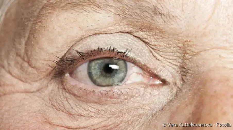
One of the most common eye diseases in old age is cataract (2), having a very high prevalence (1). Cataracts have many different triggers (3) and affects workers from all professions (6). Today, cataract surgery and appropriate follow-up treatment enable almost 90 percent of all patients (4) to regain between 50 and 100 percent (7) of their original visual performance (5).
To know more about cataracts, we have a complete study in this article.
The ICD codes for this condition are H28, H25, Q12, and H26
Read here how cataract surgery and aftercare for the eye disease are performed.
Cataract Surgery At A Glance
Cataract surgery (cataract operation) is the most frequently performed eye surgery. In the European Union alone, around 600,000 such operations are performed each year, worldwide there are more than 100 million.
The operation can be performed on an outpatient basis and usually takes less than 30 minutes. The procedure is a so-called microsurgical operation, which means that it is performed with an operating microscope. This is possible in every hospital or in the practice of an ophthalmologist.
The principle of every cataract surgery is to remove the cloudy lens and replace it with an artificial intraocular lens. As a rule, however, the entire lens is no longer removed today. Instead, the lateral and posterior lens capsules are usually left in the eye.
The intraocular lens must have exactly the same refractive power as the removed lens. The appropriate lens strength is calculated by the doctor measuring the patient’s eye length with an ultrasound device and determining the refractive power of the cornea.
After cataract surgery, the artificial lens remains in the eye for life. So it does not have to be replaced at some point.
Cataract surgery: When is it indicated?
When a cataract is operated on depends on various factors. The patient determines the time of surgery together with the attending physician.
The main factor in the decision is how much the affected person feels impaired by visual impairment in everyday and professional life. People who drive must have an eye test at regular intervals. From a certain level of visual impairment, participation in road traffic is no longer possible. The patient’s fear of eye surgery is also taken into account in the decision.
If the employer requires a certain visual performance, e.g. for pilots, professional drivers or athletes, an operation is often necessary at an early stage of the illness. The subjective perception of visual performance is not important here.
If other eye diseases are present, it is necessary to clarify beforehand with the ophthalmologist which visual acuity can probably be achieved with cataract surgery. A less good result of the operation can be expected in the following diseases:
- age-related macular degeneration (AMD)
- Retinal disease due to diabetes mellitus (diabetic retinopathy)
- Glaucoma (green star)
- Ischaemia
With advanced cataracts, visual performance can deteriorate so dramatically that blindness is imminent. In such a case, the operation should be performed even if you are afraid of the operation. Since cataract surgery is one of the most frequently performed surgeries of all, attending physicians have a lot of routine experience in it. In any case, the prospect of an improvement in visual performance outweighs the possible side effects and complications of the operation.
A congenital cataract should be operated on immediately after diagnosis. Only then is there a chance that the child can learn to see properly.
Cataract surgery: Lenses used
In cataract surgery, the patient’s own lens is replaced by an artificial lens. Today, artificial lenses are available in many different materials. They are classified according to the implantation site, the material used or according to their optical principles:
Differences in the implantation site
Depending on the implantation site, anterior chamber lenses, posterior chamber lenses and iris-supported lenses are distinguished:
In cataract surgery, anterior chamber lenses (VKL) are inserted into the anterior chamber of the eye (in front of the iris) and held there at the chamber angle with two temples. They are only used for the so-called intracapsular cataract extraction (see below). Anterior chamber lenses are now rarely used because of the risk of causing glaucoma or clouding of the cornea.
Posterior chamber lenses (HKL) are inserted into their own capsular bag, which is located behind the iris. If, as in intracapsular cataract extraction, there is no capsular bag left, the lens is attached to the iris or sclera of the eye with two sutures.
Iris worn lenses (iris clip lenses) are attached to the iris with small bows. Since the cornea is often injured in this process, such lenses are no longer used in most countries. Already implanted iris worn lenses are often replaced by posterior chamber lenses.
Material Differences In The Lenses
For cataract surgery with small incisions, intraocular lenses made of silicone or acrylic are used, as these lens materials are foldable. These artificial lenses are inserted into the capsule in a folded state, where they unfold themselves. An acrylic lens has a higher refractive index than a silicone lens and is therefore slightly thinner. The foldable silicone or acrylic lenses are used exclusively as posterior chamber lenses.
In contrast, dimensionally stable lenses made of polymethyl methacrylate (PMMA, Plexiglas) can be used as anterior chamber lenses as well as posterior chamber lenses. In this case a slightly larger incision is required for the implantation.
Differences in the optical principles
The optical principle of a lens is understood to be the characteristics that determine the new “vision” of the person concerned. Physicians distinguish between mono-focal lenses and multifocal lenses:
A mono-focal lens is similar to normal glasses. It has only one focal point, which means that the affected person can see sharply either in the distance or close up. Before the operation, the patient must decide whether he/she would prefer to live without “distance glasses” but with reading glasses after the operation or vice versa. The appropriate strength of artificial lenses is selected accordingly.
Multifocal lenses allow good visual acuity for both distance and near vision. For more than 80 percent of daily tasks, it is then no longer necessary to wear glasses. However, multifocal lenses have two disadvantages due to their special design principle: Contrasts are seen less sharply and the eye becomes more sensitive to glare.
The Procedure Of The Cataract Surgery
Cataracts usually occur on both eyes. However, only one eye is operated on first. As soon as this eye is healed, the second eye will take its turn.
A cataract operation is usually performed on an outpatient basis and under local anaesthetic. In most cases, the administration of suitable eye drops is sufficient for anaesthesia; alternatively, a local anaesthetic can be injected into the skin next to the eye to be operated on. The entire eyeball becomes painless and can no longer be moved. You can also be given a light sedative before the operation.
Normally, a cataract operation takes less than 30 minutes. Throughout the entire operation, your circulation is monitored by a blood pressure device, by measuring your oxygen saturation or by means of an ECG.
There are various methods of lens implantation to remove the lens clouding. Which one is used in each individual case depends on individual conditions and the stage of the disease.
1. Intracapsular cataract extraction (ICCE)
In this surgical method, the lens and capsule are removed from the eye. This requires a relatively large incision (eight to ten millimetres) through the cornea. The lens is then frozen with a special cold pen and removed from the eye. The artificial lens can now be inserted either in the anterior chamber (anterior chamber lens) or in the posterior chamber (posterior chamber lens). The cut is then sewn with a fine thread.
Intracapsular cataract extraction is usually only necessary at an advanced stage of the disease.
2. Extracapsular cataracts extraction (ECCE)
In extracapsular cataract extraction, the surgeon opens the anterior lens capsule with an approximately seven-millimeter long incision and removes the lens core without crushing it. The artificial lens is then inserted into the intact capsule. This surgical method is gentle on the cornea. Therefore, it is mainly used when a highly advanced cataract has already damaged the thin innermost layer of the cornea (corneal endothelium).
3. Phacoemulsification
In phacoemulsification, the cornea is opened with an incision about three millimetres wide. Then the lens core is dissolved and suctioned off with the help of ultrasound or laser. The artificial replacement lens is inserted into the intact envelope of the lens (capsular bag). It is folded, pushed through the tiny opening and unfolds in the capsular bag itself. Two semi-circular elastic temples at the edge of the lens ensure a secure fit in the capsular bag.
The tiny incision made during phacoemulsification closes itself after the operation without scarring. In the end, the surgeon only has to close the previously retracted conjunctiva again.
Thanks to the small incision it is possible with this surgical method to fit a new pair of glasses earlier than with the others and to resume the accustomed everyday life.
Cataract Surgery: What Happens Afterwards?
After the operation, the operated eye is covered with an ointment bandage. For some time you will have to stay in hospital or in the doctor’s surgery for monitoring – but as long as no complications arise, you may go home after a few hours. Subsequently, regular check-ups by the attending physician are necessary.
Please note that you must not drive a car yourself immediately after a cataract operation. So you should send someone to pick you up.
After the operation you may eat and drink light food and beverages on the same day. You can usually take your usual medication as usual, but you should discuss this with your doctor beforehand. Prior consultation is particularly advisable if you need medication for diabetes or blood-thinning medication.
As long as the operated eye is covered with a bandage and the surgical wound has not yet healed, you should take care when showering and washing that the eye does not come into contact with soap. Physical exertion, swimming, diving, cycling or sauna visits should be avoided at first. The same applies to activities that produce a lot of dirt or dust.
You can usually read and watch television again after a week.
A new pair of glasses can usually be fitted four to six weeks after cataract surgery. At an earlier stage, this is not yet useful, as the eye first has to get used to the new lens.
Cataract Surgery: Risks And Complications
Approximately 97 to 99 percent of all cataract operations are performed without complications. Nevertheless, as with any surgical procedure, there are risks:
Capsule rupture
If the posterior capsule of the lens tears during surgery, complications may occur. Behind the lens of the eye is the so-called vitreous body. It consists of a gel-like, transparent mass and presses the retina, which is located in the posterior part of the eye, against its base. If the vitreous substance escapes via a tear in the lens, a retinal detachment may occur. This risk is present in about six to eight percent of intracapsular operations; in an extracapsular operation, however, a capsule rupture hardly ever occurs.
Bacteria
Very rarely, bacteria enter the inside of the eye during intracapsular cataract surgery and lead to inflammation (endophthalmitis). The affected eye can go blind.
Bleeding
During the operation, there may be an increase in pressure in the eye, which may cause blood vessels to burst. Bleeding within the eye (intraocular) or within the capsule (intracapsular) is the result. However, they are very rare: Such bleeding occurs in less than one percent of all cataract operations.
Corneal Curvature
With the extracapsular surgical method, the incision causes a slightly stronger corneal curvature than before the operation. Usually, however, this recedes of its own accord within a few weeks.
When to see a doctor
If you notice the following signs some time after the operation, you should definitely go to an ophthalmologist:
- Visual acuity deterioration
- increased redness of the eye
- Pain in the eye
Cataract surgery: The secondary cataract
Depending on the surgical technique, 20 to 30 percent of the operated patients can develop a so-called secondary cataract (Cataracta secundaria). This usually happens more often with young people than with older patients. In the case of a post-star, the rear parts of the remaining lens capsule become cloudy. With the help of a laser or another surgical procedure (similar to cataract surgery), these cloudy parts of the lens can be quickly removed with minimal risk. The eyesight improves again afterwards.