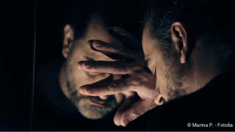
Pancreatitis: symptoms, causes, therapy
Pancreatitis: symptoms, causes, therapy
An inflammation of the pancreas (pancreatitis) can be acute or chronic. The acute inflammation is triggered by gallstones in most patients. It causes the gland to “digest itself”, so to speak. The typical symptom of acute pancreatitis is belt-shaped upper abdominal pain radiating into the back. Read more about symptoms, causes, diagnosis and treatment of pancreatitis!
Brief overview
- Definition: Acute or chronic inflammation of the pancreas (gland in the abdominal cavity that produces digestive enzymes and important hormones)
- Symptoms: Acute pancreatitis: severe pain in the upper abdomen that may radiate to the back; also gummy tummy, nausea and vomiting, fever, jaundice, bluish discoloration of the skin. Chronic pancreatitis: weight loss, digestive problems, fatigue, gait disorders, night blindness, bleeding tendency, etc.
- Causes: In acute pancreatitis, usually gallstones or alcohol consumption, less often medication, infections, etc. The trigger for chronic pancreatitis is usually regular alcohol consumption. More rare causes are metabolic diseases or genetic defects.
- Investigations: Patient interview (anamnesis), physical examination, blood test (pancreatic enzymes, calcium, gamma-GT, alkaline phosphatase, liver values etc.), stool examination, imaging procedures (ultrasound, X-ray, possibly magnetic resonance imaging, computer tomography), biliary endoscopy (ERCP)
- Treatment: Acute pancreatitis: hydration, medication, short-term dietary restrictions, followed by a light diet, possibly artificial nutrition, removal of existing gallstones; rarely surgery is necessary. Chronic pancreatitis: life-long abstinence from alcohol, intake of digestive enzymes and vitamins, possibly insulin administration, rarely surgery.
- Prognosis: Depends on the course (acute/chronic) and severity of the inflammation.
Acute pancreatitis: symptoms
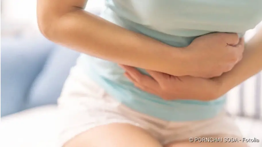
Sudden, severe pain in the upper abdomen is a common sign of acute pancreatitis. Symptoms such as a bloated stomach as well as nausea and vomiting are often added. However, the type and extent of the complaints depend on the severity of the inflammation and possible consequences. In summary, acute pancreatitis can lead to the following symptoms and complications:
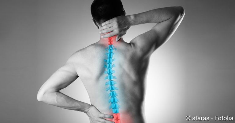
- acute, severe upper abdominal pain: Sudden upper abdominal pain that radiates in a belt-like manner to the back or sometimes in another direction and can last for several days. If gallstones are the trigger for acute pancreatitis, the pain can be colicky (i.e. it increases and decreases in waves).
- Nausea and vomiting: Upper abdominal pain in acute pancreatitis is usually accompanied by nausea and vomiting.
- Flatulence and bloated abdomen: In acute pancreatitis, the abdomen is often rubbery (“rubber belly”) and sensitive to pressure.
- Facial flushing
- Fever and weakness: Temperatures above 38°C are a defence mechanism of the body that occurs during inflammatory reactions. Often the blood pressure is also very low. Both lead to the patient feeling weak and tired.
- Circulatory problems: Released inflammatory substances cause the vessel walls to become more permeable. This makes it easier for liquid to leak into the surrounding tissue. This causes the blood pressure to drop, which can cause circulation problems. Possibly even a circulatory shock will occur!
- Water accumulation in the abdomen and lungs: As a result of acute pancreatitis, fluid may accumulate in the abdominal cavity (abdominal dropsy, ascites) or between the lungs and the chest wall (pleural effusion).
- Bluish skin discoloration: In severe acute pancreatitis, bluish-greenish spots (bruises) may appear around the navel (Cullen sign) or on the lateral flanks (Grey Turner sign). They are caused by small bleedings in the superficial fatty tissue. This symptom is considered an unfavourable sign for the further course of acute pancreatitis.
- Jaundice (icterus): In most cases the acute pancreatitis is due to a bilious disease. If this organ no longer works properly, bilirubin, a breakdown product of the red blood pigment (haemoglobin), remains in the body and is deposited in various places. First of all, there is the sclera, the white of the eye, which subsequently becomes discoloured. Later, the bilirubin is also deposited in the skin and mucous membranes, which also take on a yellow colouring as a result.
Acute pancreatitis: causes and risk factors
Acute pancreatitis is caused by gallstones in about 45 percent of cases (biliary pancreatitis): The bile (produced in the liver and temporarily stored in the gallbladder) and the digestive secretions from the pancreas (pancreatic juice) usually flow through a common duct into the small intestine. If this passage is blocked by gallstones, the pancreatic juice backs up into the pancreas (and the bile towards the liver). The digestive enzymes contained in the accumulated pancreatic juice then become active in the pancreas instead of in the intestine as intended. They attack the glandular tissue so that the pancreas digests itself to a certain extent (proteolytic autodigestion). The damaged tissue calls the immune system into action and an inflammatory reaction occurs.
Acute pancreatitis is usually caused by a disease of the bile ducts, such as gallstones, which block the ducts for bile and pancreatic secretions.
Alcohol is the second most common cause of acute pancreatitis. He is responsible for about 35 percent of all cases of illness. Alcohol attacks the pancreatic tissue directly and thus causes an inflammatory reaction.
In order to get acute pancreatitis, it is not necessarily necessary to be drunk. Some people already react to a relatively moderate amount of alcohol with pancreatitis.
Rarer causes of acute pancreatitis (about 15 percent of all cases) are
- Viral infections such as mumps, HIV, viral hepatitis
- certain drugs such as dehydrating agents, blood pressure reducers, hormones
- highly elevated calcium level, e.g. in cases of parathyroid overactivity
- highly elevated blood fat values (> 1000 mg/dl)
- Endoscopy of the bile duct system (ERCP), abdominal injury or surgery
- Inheritance (hereditary pancratitis)
- special anatomical features
In about 15 percent of those affected, no cause for the pancreatitis can be found. Doctors then call this idiopathic pancreatitis.
Chronic pancreatitis
In contrast to acute pancreatitis, chronic pancreatitis is a recurrent inflammation of the pancreas. The trigger is regular alcohol consumption in 70 to 80 percent of cases. People react differently to alcohol: sometimes even small amounts of alcohol are enough for a chronic inflammation of the pancreas.
The disease often begins insidiously with mild to moderate symptoms. Belt-shaped pain in the upper abdomen is a typical symptom – as in acute pancreatitis. They often stay open after dinner. Other common signs of chronic pancreatitis include weight loss, digestive problems, nausea, vomiting and fatty, shiny, foul-smelling stools (fatty stools).
You can read more about the causes, symptoms, treatment and consequences of a chronically inflamed pancreas under Chronic Pancreatitis.
Acute pancreatitis: examinations and diagnosis
If you develop possible signs of (acute) pancreatitis, you should go to your family doctor or directly to a specialist in internal medicine and gastroenterology. If the complaints occur outside office hours, it is advisable to visit a hospital. Especially in case of severe symptoms you should not hesitate to seek medical help! Acute pancreatitis in particular can be not only very painful, but under certain circumstances also life-threatening.
Collection of the medical history
First, the doctor will ask you about your symptoms in detail. He will also inquire about possible triggers for pancreatitis. Typical questions during this anamnesis interview are:
- Do you have a fever or are you feeling sick?
- Did the symptoms appear suddenly? Or do you appear on certain occasions?
- Are you familiar with gallstones?
- Have you taken any medication and, if so, what kind?
- Did you drink a lot of alcohol before the symptoms appeared or do you drink alcohol regularly?
- Did a blood test show elevated blood lipid or calcium levels?
Physical examination
The interview is followed by a physical examination. The doctor will ask you to clear your stomach. This allows the doctor to check whether you have an elastic and tight “rubber belly” – a common sign of acute pancreatitis. In addition, the stomach is often very sensitive to pain. Patients therefore often bend their legs in order to alleviate the pain somewhat.
During the physical examination, the doctor also looks for bruises in the lateral flanks and around the navel and checks whether the white of the eyes and the skin have turned yellow.
Blood tests
Certain blood values help to rule out or confirm (acute) pancreatitis: For example, elevated levels of the pancreatic enzymes lipase and alpha-amylase are detectable in the blood when pancreatitis occurs. However, elevated enzyme levels can also have other causes, so they are not proof of pancreatitis.
Other blood values that are determined when pancreatitis is suspected are blood sugar, kidney and liver values. The calcium level is also measured: Increased calcium levels can be the cause of acute pancreatitis.
Elevated levels of gamma-GT and alkaline phosphatase (AP) may indicate bile stasis.
Furthermore, the doctor can measure the inflammation values like the C-reactive protein (CRP). If it is elevated, this generally indicates an inflammation in the body. The CRP value is also suitable for monitoring the course of acute pancreatitis.
Imaging methods
In order to be able to diagnose pancreatitis with certainty, the doctor must examine the abdominal cavity with an imaging procedure. The easiest and fastest method is an ultrasound examination (sonography). This often makes it possible to detect gallstones, which are the most common cause of acute pancreatitis. In addition, the doctor can use ultrasound to assess the extent of the inflammation and detect disease-related changes. This can include swelling of the pancreas, dead tissue and water retention in the abdomen and lungs.
If the ultrasound findings are inconclusive, magnetic resonance imaging (MRI) or computed tomography (CT) can provide clarity. Both methods provide very detailed images of the examined tissue. They are also used in cases of severe pancreatitis.
By means of an X-ray examination, the doctor can check the function of the lungs and intestines. Water accumulation between the lungs and chest wall and in the abdominal cavity can be easily detected on the X-ray. Any air bubbles in a paralysed intestine are also visible.
Endoscopic examination
If bile ducts are suspected to be blocked by gallstones or a tumour in the area of the bile ducts, the doctor can perform a biliary endoscopy: More precisely, it is a mirror image of the pancreatic and bile ducts and the gall bladder. The medical term for this endoscopic examination is endoscopic retrograde cholangio-pancreaticography (ERCP).
With ERCP, gallstones that are detected can usually be removed immediately.
Stool test
If chronic pancreatitis is suspected, the doctor can send a stool sample from the patient to the laboratory for analysis. There the content of the pancreatic enzyme elastase is measured, which is normally excreted unchanged with the stool. In the event of tissue damage (for example as a result of chronic pancreatitis) less elastase reaches the small intestine and stool. Then the elastase concentration in the faeces is unusually low.
Since some diseases can cause similar symptoms to pancreatitis, the doctor must exclude them in the course of the diagnosis. These diseases include heart attack, appendicitis, biliary and renal colic, tubal pregnancy and pulmonary embolism.
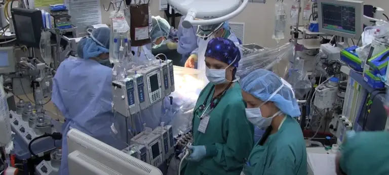
Acute pancreatitis: treatment
Acute pancreatitis is potentially life-threatening. Patients must therefore be treated in hospital, in severe cases even in intensive care. An adequate fluid intake and appropriate pain therapy are important components of the treatment. In addition, the diet must be adjusted in the event of pancreatitis.
Pancreatitis – fluid administration
In patients with pancreatitis, a lot of fluid from the blood vessels seeps into the tissue. This causes a lack of fluid in the blood vessels. The blood pressure drops, possibly to the point of circulatory failure due to shock. The patient therefore receives sufficient fluid by infusion. If they also lack blood salts (electrolytes), these are also supplied with the infusion.
Pancreatitis – Medications
Painkillers and antispasmodic drugs help against the strong, often cramping pain. Patients with severe acute pancreatitis often receive antibiotics as well: The aim is to prevent or combat a bacterial infection of the inflamed pancreas at an early stage. In some cases, acute pancreatitis can lead to the formation of blood clots (thromboses), which is why doctors inject the anticoagulant heparin as a precaution.
Pancreatitis – Nutrition
To relieve the pancreas, patients should not eat for a few days. This is especially true when nausea and vomiting occur. Especially in cases of severe acute pancreatitis and complications, patients are supplied with the necessary nutrients via infusions (parenteral nutrition). Sometimes a small intestinal tube is inserted later: The doctor carefully passes a thin tube through the patient’s nose or mouth, through the oesophagus and stomach, and into the small intestine. Via this probe, nutrients can be conducted directly into the intestine.
As soon as the patient is allowed to eat food again, a light bland diet (rusk, rice gruel, tea etc.) is started. Fat-containing food is only allowed again after two to three weeks and then for the time being only in small quantities. Alcohol is taboo. More detailed tips on nutrition in the case of pancreatitis are given by the doctor treating the patient. He may also recommend the patient to temporarily take enzyme preparations that support digestion.
Gallstone removal
If gallstones are the cause of acute pancreatitis (biliary pancreatitis), they are usually removed endoscopically using ERCP. Larger stones are first crushed with shock waves (shock wave therapy).
Operation
In the case of a severe acute pancreatitis, surgery is sometimes necessary. Especially if a lot of pancreatic tissue has died off due to inflammation (necrotizing pancreatitis), it must be cut out.
Acute pancreatitis: course and prognosis
As long as no complications occur, the prognosis for acute pancreatitis is usually good. With proper treatment, about 80 percent of patients are healthy again after about one to two weeks. In about 20 percent of cases, however, serious consequences develop. The healing process can then take weeks or months. Severe acute pancreatitis kills about 15 percent of those affected.
Indicator Ranson-Score
In order to better assess the prognosis for acute pancreatitis in individual cases, physicians use the so-called Ranson Score. Various clinical parameters and laboratory values are assessed within the first 48 hours after the patient is admitted to the clinic:
| On admission to hospital | ||
| Age | > 55 years | 1 point |
| White blood cells (leukocytes) | > 16000 per mm3 | 1 point |
| Lactate dehydrogenase (LDH) | > 350 U/litre | 1 point |
| Aspartate aminotransferase (ASAT) | > 250 U/litre | 1 point |
| Glucose | > 11.1 millimoles per litre or > 200 milligrams per decilitre | 1 point |
| 48 hours after admission to hospital | ||
| Haematocrit waste | > ten percent | 1 point |
| Urea Increase | > 1.8 millimol per litre | 1 point |
| Calcium level in serum | < 2 millimoles per litre | 1 point |
| Arterial oxygen partial pressure | < 60 mm Hg | 1 point |
| Deficiency in blood bases | > 4 mEql | 1 point |
| Loss of fluid from the vascular system to the tissue | > 6 litres in 48 hours | 1 point |
A point is awarded in the Ranson Score for each applicable criterion. The higher the score, the greater the risk that acute pancreatitis will be fatal (mortality prognosis).
Possible complications
If acute pancreatitis is not treated, it can cause complications anywhere in the body:
Volume deficiency shock (hypovolemic shock): The blood vessels become more permeable as a result of the inflammation. If as a result too much fluid leaks into the surrounding tissue, a dangerous lack of volume in the vascular system develops – the blood pressure can drop so much that the organs can no longer be supplied with sufficient oxygen. The result is hypovolemic shock.
Bowel paralysis: Any inflammation in the service room can disturb the normal movements of the bowel. These can even come to a complete standstill, so that the food pulp can no longer be transported. Then you have a life-threatening intestinal paralysis (paralytic intestinal obstruction)! Possible signs are severe pain and flatulence. In extreme cases the patient vomits stool. An intestinal obstruction must always be treated as quickly as possible!
Pancreatic pseudocysts: After an acute pancreatitis has subsided, fluid-filled cavities can form in the pancreas, which are surrounded by collagen fibres and wound healing tissue. They are called pancreatic pseudocysts. Often they remain small, do not cause any symptoms and disappear on their own within a few weeks. But sometimes they also cause complaints such as indigestion or a feeling of fullness. In addition, larger pseudocysts are at risk of rupturing and bleeding or becoming infected and forming an abscess. In order to prevent this from happening, the liquid in larger pseudocysts is sucked out from outside via a hollow needle (drainage). Sometimes an operation is also necessary.
Necrotizing infectious pancreatitis: Rarely does acute pancreatitis develop into necrotizing infectious pancreatitis. This means: glandular tissue dies off as a result of inflammation (necrosis) and the dead tissue becomes infected with bacteria. This dangerous complication can lead to a so-called SIRS (systematic inflammatory response syndrome): It is characterized by a spread of the inflammation to the whole body. Possible signs are high fever (but sometimes also a lowered temperature), fast pulse and rapid breathing. There is a risk of organ failure!
Acute pancreatitis: prevention
For those affected, it usually seems as if the acute pancreatitis came out of nowhere. In fact, however, such a disease often takes a long time to develop through a corresponding lifestyle. To prevent acute pancreatitis, you should therefore
- eat low-fat food
- drink little or no alcohol
- Diseases such as elevated blood lipid levels or parathyroid hyperthyroidism
Especially people who have already had an acute pancreatitis should follow these tips!