Spinal stenosis: causes, symptoms, therapy
Spinal canal stenosis: causes, symptoms, therapy
In spinal canal stenosis (spinal stenosis, spinal stenosis, spinal canal stenosis), the canal in the spine through which the spinal cord passes is narrowed. The resulting pressure on the spinal cord, nerves and blood vessels can cause back pain and permanent nerve damage. In most cases, a spinal canal narrowing occurs due to aging processes. Here you can read interesting facts about the causes, symptoms and treatment options of spinal canal stenosis.
Brief overview
- Causes: Mostly wear (degeneration) of the spine. Less frequently congenital constrictions (hollow back, malformations of the spine), spinal surgery, disc protrusion or prolapse, hormonal changes, bone diseases such as Paget’s disease.
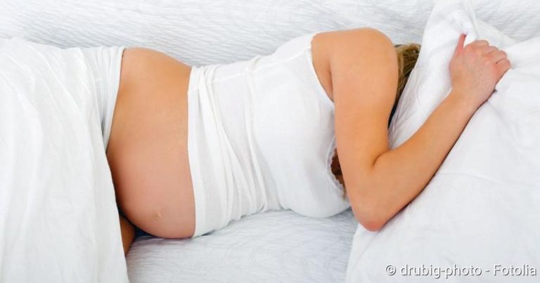
- Symptoms: Often asymptomatic at first. Later, often unspecific complaints such as back pain radiating into the legs (lumboischialgia), reduced mobility of the lumbar spine and muscle tension in the lower back. In cases of severe spinal stenosis sensory disturbances and weakness in the legs, limping (claudatio spinalis), bladder and/or rectum disorders, disturbed sexual function. Very rarely paraplegic syndrome with paralysis of the legs as well as problems with bowel movements and/or urination.
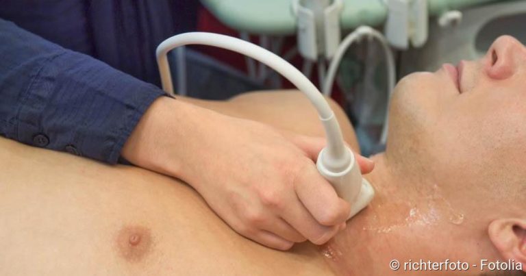
- Diagnosis: On the basis of characteristic symptoms in combination with imaging techniques (magnetic resonance imaging, computer tomography).
- Treatment: Mostly conservative therapy with a combination of physiotherapy, back school, heat therapy, electrotherapy, support corset (orthosis), pain management training and pain therapy. An operation is rarely necessary to relieve the pressure on the spinal canal.
- Course and prognosis: Even without therapy the course is usually very slow. With consistent conservative therapy, the typical complaints can usually be well treated and alleviated.
Spinal stenosis: causes
The most frequent cause of spinal stenosis is wear and tear (degeneration) of the spine: over time, the intervertebral discs between the vertebral bodies lose fluid. This makes them flatter and thus less able to cushion the pressure caused by movement – the vertebral bodies are thus subjected to greater loads and then press on the spinal canal.
Due to the shrunken intervertebral discs, the ligaments along the spine are also less taut and lose their elasticity. Thus the entire spinal column structure can become latently unstable. The vertebral bodies can shift against each other (spondylolisthesis) and compress the spinal canal.
A well-trained back musculature then stabilizes the spine so that one is free of complaints despite spinal canal stenosis. In contrast, patients with poorly developed back muscles often develop typical stenosis symptoms. This is because if the muscles cannot support the unstable spine, the body forms new bone structures on the vertebrae to stabilize the spine. These newly formed bony structures are called osteophytes. They can not only aggravate a spinal canal narrowing but also cause it.
In spinal stenosis, the spinal canal through which the spinal cord passes narrows. In most cases, wear and tear of the vertebral bodies or intervertebral discs is the cause.
Arthrosis of the vertebral joints (facet joints) can also lead to new bony formations and thus promote spinal stenosis.
Rarer causes of spinal stenosis are
- congenital malformations such as a strong hollow back, spondylolisthesis, chondrodystrophy (disorders in the transformation of cartilage into bone tissue at embryonic age). In such cases, the complaints already occur at the age of 30 to 40 years.
- Spinal surgery (the excessive formation of scar tissue can narrow the spinal canal)
- Injuries to the vertebral bodies
- Protrusions or prolapse of disc material into the spinal canal
- hormonal changes that affect the bone substance and the stability of the vertebral bodies (e.g. Cushing’s disease)
- Bone diseases (such as Paget’s disease): This involves a locally limited remodelling and cultivation of bones. In this disease, spinal canal stenosis occurs more frequently in the lumbar than in the cervical spine.
- spinal canal narrowed from birth of unknown cause (idiopathic spinal canal stenosis)
Spinal canal stenosis: symptoms
A spinal canal stenosis usually occurs in the lumbar spine (lumbar spinal canal stenosis). It does not necessarily lead to complaints. These only occur when the spinal canal is so narrowed that nerves or blood vessels are compressed. Which complaints these are, when and how severe they are, depends on several factors. These include the severity of the disease, the patient’s posture and the extent of physical strain.
At the beginning of the disease the symptoms are not very characteristic and diverse. These non-specific complaints include
- Back pain in the lumbar region (lumbago), which usually radiates into the legs on one side (lumboischialgia)
- reduced mobility in the lumbar spine
- Muscle tension in the lumbar spine
If the stenosis progresses further, the following complaints are possible:
- Sensory disturbances in the legs
- Sensations in the legs, e.g. burning, formication, feeling cold, cotton wool feeling under the feet
- Weakness in the leg muscles
- limping due to pain (Claudicatio spinalis)
- Bladder and/or rectal problems (problems with bowel movement and urination or incontinence)
- dysfunctional sexual function
It is typical for the disease that the symptoms usually improve significantly when sitting or in other positions where the trunk is bent forward (for example when cycling, bending down and walking uphill).
Limping in spinal canal stenosis (claudicatio spinalis) must be distinguished from temporary limping due to circulatory disorders in “window dressing” (pAVK). The latter is called Claudicatio intermittens.
Very rarely, spinal canal stenosis leads to a so-called paraplegic syndrome: Both legs are paralyzed, and problems with bowel movement and urination occur.
Sometimes the spinal canal narrowing does not affect the lumbar spine but the cervical spine (cervical spinal canal stenosis). Those affected often have neck pain that radiates into the arms. Over time, sensory disturbances in the legs, rectal and bladder problems may develop.
Spinal stenosis: treatment
In most cases, spinal stenosis can be treated well with conservative therapy methods. Only rarely (in very severe cases) is a surgical intervention necessary.
Conservative treatment
The conservative forms of therapy for spinal canal stenosis include
- Physiotherapy (movement therapy, baths, muscle relaxing treatments etc.) to relieve and stabilize the spine
- Heat therapy to relax the back muscles
- Electrotherapy for pain treatment and muscle relaxation
- Support corsets (orthoses) to relieve the spinal column
- Back school (targeted strengthening training for the back and abdominal muscles, tips for back-friendly postures, behavioural tips)
- Psychological pain management training
- Pain therapy
Usually several of the above measures are combined. One then speaks of a modular therapy concept.
Medication
An effective pain treatment is a cornerstone of conservative stenosis therapy. Depending on the intensity of the pain, different agents are used. For mild pain, non-opioid painkillers such as ibuprofen, paracetamol or diclofenac can help. They have an analgesic and anti-inflammatory effect (the latter, however, is only very slight with paracetamol). For moderate pain, light opioids are used, often in combination with light (non-opioid) painkillers. Severe pain is treated with strong opioids.
Some painkillers can irritate the stomach lining if taken for a long time. That is why doctors often prescribe so-called proton pump inhibitors to accompany the treatment. As “stomach protection”, these drugs ensure that the body produces less stomach acid.
In addition to the classic painkillers, the doctor may prescribe mild antidepressants. In small doses they can help with chronic pain.
Sometimes muscle-relaxing medication can also help with spinal stenosis. If the pain is very severe, a high-dose cortisone therapy may be considered: cortisone causes the soft tissues that press on the spinal canal to swell. So there is a little more space in the canal again.
The various active ingredients with analgesic, anti-inflammatory, local anaesthetic and/or decongestant effects can often be administered not only by mouth (as tablets, capsules etc.). In many cases, they can also be injected directly into the affected area of the spinal canal stenosis. This injection therapy is particularly indicated when a nerve root is severely inflamed. The combination of anti-inflammatory agents such as cortisone and painkillers fights the inflammation and at the same time alleviates the pain-related complaints. If, on the other hand, the pain emanates from the small vertebral joints, local injections with painkillers can help.
In studies on injection therapy, patients received ineffective substances (placebo) instead of real medication, often simple table salt. Despite this sham treatment many patients had less pain afterwards. The researchers discovered that the placebo injections released the body’s own “painkillers” (endorphins).
Operation
Almost all patients with spinal canal stenosis are helped by conservative therapy. An operation is rarely necessary – usually when important nerves fail. In addition, surgery can be performed if conservative treatment fails or the patient has a high level of suffering and is significantly restricted in his or her everyday life.
The surgical procedure always aims to relieve the region where the spinal cord is squeezed. Various methods are available for this purpose:
- Pressure relief (decompression) of the constricted nerves is considered the method of choice. For this purpose, the vertebral arch is removed at the stenosis site on one or both sides together with the spinous process (hemi/laminectomy). Sometimes only parts of the vertebral arch are removed (micro-decompression).
- Fusion (Spondylodesis): Individual vertebrae are connected and stiffened by material from the iliac crest or by screws. This prevents them from sliding into each other and constricting the spinal canal.
- Interspinous implants connect the spinous processes of the vertebral bodies and thus prevent the spinal column in the affected area from tilting forward or backward.
The doctor decides which method is most suitable in each individual case. All three procedures can usually be performed minimally invasive or microsurgically. This means that the physician does not have to make a large cut to reach the affected region. Several small incisions are sufficient for the surgeon to introduce a tiny camera with light source and the fine surgical instruments.
Every operation involves certain risks. In addition, the “skin” around the spinal cord can be damaged, causing spinal fluid to leak out (liquor fistula). Before operating on a spinal canal stenosis, the physician will therefore carefully weigh up the expected benefits and potential risks against each other.
Spinal canal stenosis: examinations and diagnosis
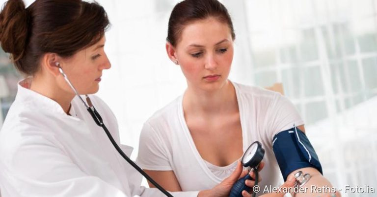
In the initial consultation (anamnesis), the doctor will ask the patient in detail about his or her complaints and known pre-existing or underlying diseases (herniated disc, arthrosis, osteoporosis, etc.). This is followed by a physical examination: among other things, the doctor may ask the patient to bend the upper body far back and then forward. If spinal stenosis is present, the back hurts when leaning back, while the symptoms disappear when the trunk is bent.
Imaging techniques provide further information. Here, experts recommend above all magnetic resonance imaging (MRI) without contrast medium. It can show nerves, intervertebral discs, blood vessels and other structures in very detailed layered images.
Alternatively, the spine can be visualized by computer tomography with a contrast medium. However, this so-called myelo-CT exposes the patient to a certain amount of radiation.
Not every narrowing of the spinal canal that is visible on MRI or other imaging procedures actually causes symptoms!
In addition, the doctor can X-ray the patient while standing and in certain postures (functional images).
Electrophysiological examinations can also be used to clarify spinal canal stenosis. These include electromyography (EMG) and so-called evoked potentials. These methods help to assess the function of nerves.
Spinal canal stenosis: course and prognosis
Even if it is not treated, spinal stenosis usually progresses very slowly. Depending on the cause, the disease can also progress very differently. The pain caused by the pressure on the nerve tracts can remain constant, decrease with certain movements or over time, or come and go constantly. Sometimes the symptoms even decrease with age, when the spine becomes less mobile. This is because the nerves are then irritated less often, so that movement-related pain occurs less frequently.
In some cases, however, spinal canal stenosis is acute: if, for example, intervertebral disc tissue shifts (protrusion, prolapse), capsular swelling occurs in osteoarthritis, or fluid accumulates near the nerve tracts, the symptoms of spinal canal stenosis can suddenly worsen. One side of the body is often particularly affected.
Overall, in most cases spinal stenosis can be treated well with conservative therapy methods, so that the affected persons can live relatively painlessly.
Sources And Guidelines
- Guideline “Lumbar Spinal Canal Stenosis” of the European Society for Orthopaedics and Orthopaedic Surgery and the Professional Association of Orthopaedic Physicians (2002)