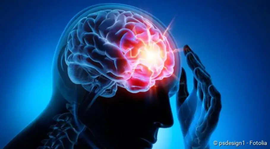
Stroke (apoplexy): warning signs, causes, therapy
Stroke (apoplexy): warning signs, causes, therapy
Stroke: short overview
- What is a stroke? A sudden circulatory problem in the brain
- Important symptoms: acute muscle weakness, feelings of paralysis and numbness in one half of the body, sudden visual and speech disturbances, acute and very severe headaches, acute dizziness, speech disorders, etc.
- Causes: Reduced blood flow in the brain, usually due to a blood clot (ischemic stroke), less frequently due to a cerebral haemorrhage (haemorrhagic stroke)
- Stroke Test (FAST Test): Ask the patient to smile (F for face), lift both arms simultaneously (A for arms) and repeat a simple sentence (S for speech). If he has problems with this, it is likely that he has had a stroke and you should quickly alert the emergency doctor (T as in time).
- First aid: Call an emergency doctor (Tel. 112), calm the patient, loosen constricting clothing, elevate the upper body (if patient is conscious), stable lateral position (if unconscious), resuscitation (if no pulse/respiration can be detected)
- Treatment: Stabilisation and monitoring of vital functions, further measures depending on the cause of the stroke (removal of the blood clot by medication or by catheter, surgery in case of extensive cerebral haemorrhage etc.), treatment of complications (epileptic seizures, increased cerebral pressure etc.)
Stroke: Description
A stroke is a sudden circulatory disturbance in the brain. It is also called apoplectic or apoplexy, stroke, brain insult, apoplectic insult or cerebral insult.
FAQ
The acute circulatory disorder of the brain results in the brain cells receiving too little oxygen and nutrients. It kills them. Losses of brain function can be the result, such as feelings of numbness, paralysis, speech or vision problems. If treated quickly, they can sometimes recede; in other cases they remain permanently. A severe stroke can also be fatal.
Causes: In addition to smoking, for example, increased blood pressure, raised cholesterol levels or diabetes. A doctor can measure this and decide whether the risk can be reduced through lifestyle measures (more conscious nutrition, weight control, regular exercise) or whether medication is necessary. A special variant of stroke is caused by atrial fibrillation. If your pulse is irregular, you must also see a doctor!
Nothing indicates the risk of a stroke as clearly as the fact that one has already occurred. The next stroke will not be long in coming unless you take countermeasures. So yes: you should definitely know if you have ever had a stroke. In case of doubt, an MRI examination of the head must be performed, which reveals areas of the brain that have died due to a stroke.
Stroke: Frequency
Affected by strokes, are normally older people. As their proportion in the population is steadily increasing, the number of stroke patients is also likely to rise, experts believe.
Anyone who has already had a stroke bears an increased risk of another stroke. For example, about 40 out of 100 people who have already suffered a stroke will have another one within ten years. The risk of further cardiovascular diseases (such as heart attack) is also increased in stroke patients.
Stroke in children
Although a stroke usually affects older people, it can also occur at a young age. Even unborn children in the womb can suffer a stroke. Possible causes include, for example, coagulation disorders, heart and vascular diseases. Sometimes an infectious disease also triggers a stroke in children.
The diagnosis of “stroke” is, nevertheless, more difficult to make in children. The reason is that brain maturation is not yet complete and therefore a stroke in children often does not become apparent until months or years later. For example, hemiplegia in newborns only becomes apparent after about six months.
Stroke: Symptoms
The stroke symptoms depend on which brain region is affected and how severe the stroke is. Very often, acute weakness, numbness and paralysis appear on one side of the body. This can be recognized, for example, by the fact that the corner of the mouth and the eyelid of one side hang down and/or the patient can no longer move one arm. The left side of the body is affected if the stroke occurs in the right side of the brain and vice versa. If the patient is completely paralyzed, this indicates a stroke in the brain stem.
Sudden visual disturbances are also common stroke symptoms: Those affected report, for example, that they can only see blurred or perceive double images. A sudden, temporary loss of vision in one eye can also indicate a stroke. Due to the acute visual disturbances it can happen that the affected person falls or – while driving a car – causes an accident.
An acute speech disorder can also be a sign of a stroke: Some patients suddenly speak in a blurred or slurred way, twist letters or cannot speak at all. Often stroke patients can no longer understand what is being said to them. This is known as speech comprehension disorder.
Other possible signs of a stroke can be sudden dizziness and very severe headaches.
You can read more about the signs and symptoms of a stroke in the article Stroke: Symptoms.
Transitory ischemic attack (TIA) – the “mini-stroke”
The term “Transitory Ischemic Attack” (TIA for short) describes a temporary circulatory disorder in the brain. It is an early warning sign of a stroke and is sometimes called a “mini stroke”.
TIA is usually caused by tiny blood clots that temporarily impair the blood flow in a cerebral vessel. The affected person notices this, for example, through temporary speech or vision problems. Sometimes weakness, paralysis or numbness may occur for a short time in one half of the body. A temporary confusion or disturbance of consciousness may also occur.
Such TIA symptoms always appear suddenly and disappear again after minutes or a few hours. Nevertheless, you should consult a doctor immediately: If the right therapy is initiated quickly, a “real” stroke can often be prevented.
You can read everything important about the “mini stroke” in the article Transitory ischemic attack.
Stroke: causes and risk factors
Physicians distinguish between different causes of stroke: The two most common are reduced blood flow (ischemic stroke) and cerebral haemorrhage (hemorrhagic stroke). In rare cases, other causes of stroke can also be identified.
Cause of stroke no. 1: reduced blood flow
Acute reduced or deficient blood flow (ischemia) in certain regions of the brain is the most common cause of all strokes. It is responsible for about 80 percent of all cases of stroke. Doctors refer to this as an ischemic stroke or brain attack.
There can be various reasons why a lack of blood supply to certain regions of the brain occurs. The most important ones are:
- Blood clot: A blood clot can block a cerebral vessel and thus prevent the blood and oxygen supply of a brain region. The clot has often formed in the heart (for example in atrial fibrillation) or in a “calcified” carotid artery and was washed into the brain with the bloodstream.
- “Vascular calcification” (arteriosclerosis): Brain vessels or vessels supplying the brain in the neck (like the carotid artery) can “calcify”: Deposits on the inner wall constrict a vessel more and more or even close it completely. The brain area to be supplied then contains too little blood and oxygen.
An ischemic stroke in the brain stem can have particularly serious consequences (brainstem infarction). This is where vital brain centres are located, which are responsible for controlling breathing, circulation and consciousness. An example of a brainstem infarction is basilar thrombosis, i.e. the occlusion of the arteria basilaris in the brainstem: in severe cases it causes complete paralysis of all extremities (tetraparesis) and coma or leads directly to death.
Stroke Cause No. 2: Cerebral hemorrhage
Bleeding in the head is the cause of about 20 percent of all strokes. Stroke caused by such a cerebral hemorrhage is also called a hemorrhagic stroke. The bleeding can occur in different places:
- Bleeding in the brain: Suddenly, a vessel directly in the brain bursts and blood leaks into the surrounding brain tissue. The trigger for this so-called intracerebral bleeding is usually high blood pressure. Other diseases, drug abuse and the rupture of a congenital vascular malformation (such as aneurysm) in the brain can also cause bleeding in the brain. Sometimes the cause remains unexplained.
- Bleeding between the meninges: Here, the stroke is caused by a bleeding in the so-called subarachnoid space: This is the gap-shaped space filled with cerebral fluid between the middle meninges (arachnoidea) and the inner meninges (pia mater). The cause of such a subarachnoid hemorrhage is usually a spontaneously ruptured aneurysm (congenital vascular malformation with bulging of the vessel wall).
Rare causes of strokes
Stroke can have other causes than reduced blood flow or cerebral haemorrhage, especially in younger people. In some patients, for example, the stroke is caused by inflammation of the vessel walls (vasculitis). Such vascular inflammations occur in the context of autoimmune diseases such as giant cell arteritis, Takayasu arteritis, Behcet’s disease and systemic lupus erythematosus.
Other rare causes of stroke are, for example, fat and air embolisms: in these cases, fat droplets or air that has entered the brain block a cerebral vessel, resulting in a cerebral infarction. A fat embolism can occur in severe bone fractures when fatty bone marrow is washed into the blood. An air embolism can occur as a very rare complication of open heart, chest or neck surgery.
Congenital coagulation disorders and the formation of blood clots in the veins are also among the rare causes of stroke.
Stroke: Risk factors
Stroke doesn’t happen out of nowhere. Various factors can contribute to its development. Some of these stroke risk factors cannot be influenced. This includes age: the risk of a stroke increases with the years of life. A genetic predisposition for a stroke cannot be influenced either.
There are also many risk factors that can be reduced in a targeted manner. This includes, for example, high blood pressure (hypertension): it leads to “vascular calcification” (arteriosclerosis), i.e. deposits form on the inner wall of the vessels. This causes the vessels to become increasingly narrow. This is conducive to a stroke. The following applies: The more severe the high blood pressure, the more likely a stroke is.
Smoking is also an avoidable risk factor for a stroke: The more cigarettes someone smokes per day and the more years the smoking career has lasted, the higher the risk of stroke. There are several reasons for this:
Among other things, smoking promotes vascular calcification (arteriosclerosis) and lipid metabolism disorders – both are further risk factors for a stroke. Smoking also causes the blood vessels to constrict. The resulting rise in blood pressure promotes a stroke.
Smoking also reduces the amount of oxygen that can be transported by the red blood cells (erythrocytes). The tissues and organs receive less oxygen as a result, as does the brain. This then signals the bone marrow to produce more red blood cells for oxygen transport. However, the blood is “thickened” by the increase in erythrocytes. This makes it flow less easily through the narrowed vessels.
Last but not least, smoking increases the blood’s readiness to clot – especially because the blood platelets become stickier. This causes blood clots to form more easily, which can clog a vessel. If this happens in the brain, it results in an ischemic stroke.
It is therefore worthwhile to stop smoking. Just five years after stopping smoking, someone is at the same risk of having a stroke as people who have never smoked.
Other important risk factors for a stroke are:
- Alcohol: High alcohol consumption – whether regular or occasional – increases the risk of a stroke. In particular, the risk of a brain haemorrhage increases. In addition, regular alcohol consumption poses other health risks (such as addictive potential, increased risk of cancer).
- Overweight: Being overweight increases the risk of many different diseases. These include diabetes, high blood pressure and strokes.
- Lack of exercise: Possible consequences are overweight and high blood pressure. Both of these factors favour a stroke.
- Fat metabolism disorders: LDL cholesterol (“bad” cholesterol) and other blood fats are part of the deposits that form on the inner walls of blood vessels in arteriosclerosis. High blood fat values (such as high cholesterol levels) therefore increase the risk of stroke via arteriosclerosis.
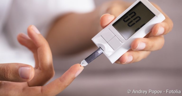
- Diabetes: Diabetes mellitus damages the walls of blood vessels, causing them to thicken. It’s affecting the blood flow. Diabetes also exacerbates existing arteriosclerosis. Overall, diabetics have a two to three times higher risk of stroke than people who are not diabetic.
- Atrial fibrillation: This cardiac dysrhythmia increases the risk of stroke because blood clots easily form in the heart. These can – carried along by the bloodstream – block a vessel in the brain (ischemic stroke). This risk is even greater if there are also other heart diseases such as coronary heart disease (CHD) or cardiac insufficiency.
- other cardiovascular diseases: Other cardiovascular diseases such as smoker’s leg (pAVK) and “impotence” (erectile dysfunction) also increase the risk of stroke.
- narrowed carotid artery (carotid stenosis): This is usually due to vascular calcification (arteriosclerosis) and often causes no symptoms for a long time. A possible early symptom is TIA (transient ischemic attack). Whether symptom-free or not – carotid stenosis increases the risk of an ischemic stroke (cerebral infarction).
- Aura migraine: Stroke caused by reduced blood flow is common in people who suffer from migraine with aura. Headaches are preceded by neurological symptoms such as impaired vision or sensation. The exact connection between aura migraine and stroke is not yet known. Women are particularly affected.
- Hormone preparations for women: Taking the contraceptive pill increases the risk of stroke. This is especially true for women with other risk factors such as high blood pressure, smoking, overweight or aura migraine. Taking hormone preparations during menopause (hormone replacement therapy, HET) also increases the risk of a stroke.
Childhood stroke: causes
Stroke in children is rare, but it happens. While lifestyle factors and diseases of civilisation (smoking, arteriosclerosis etc.) are considered the main cause of a stroke in adults, children have other causes of stroke. These include, for example, an inherited tendency to form clots, diseases of the red blood cells (such as sickle cell anaemia) and connective tissue diseases (such as Fabry’s disease). Autoimmune diseases of the blood vessels and heart disease are also possible causes of stroke in children.
Stroke: examinations and diagnosis
Whether severe or mild stroke – every stroke is an emergency! If you suspect something, you should call the emergency doctor immediately! With the FAST-Test you can easily and quickly check a suspected stroke. The FAST test helps to determine whether someone has suffered a stroke. The stroke test works as follows:
- F as in “face”: Ask the patient to smile. If the face is contorted on one side, this indicates hemiplegia as a result of a stroke.
- A as in “arms”: Ask the patient to stretch their arms forward at the same time while turning their palms upwards. If he has problems with this, it is probably due to incomplete paralysis of one half of the body as a result of a stroke.
- S for “speech”: Ask the patient to repeat a simple sentence. If he is not able to do this or if his voice sounds washed out, there is probably a speech disorder due to a stroke.
- T as in “time”: Call an ambulance immediately!
The emergency doctor will check the patient’s consciousness, blood pressure and heart rate, among other things, on site. If he is conscious, the doctor can ask him about what happened and any symptoms that occur (such as impaired vision, numbness or paralysis).
After admission to hospital, a neurologist is the responsible specialist if a stroke is suspected. He’s doing a neurological exam. In doing so, he checks, for example, the patient’s coordination, speech, vision, touch sensitivity and reflexes.
As a rule, computer tomography of the head (cranial computer tomography, cCT) is also performed immediately. The examination is often supplemented by a vascular imaging (CT angiography) or blood flow measurement (CT perfusion). The images from inside the skull show whether a vascular occlusion or a cerebral haemorrhage is responsible for the stroke. It is also possible to determine its location and extent.
Sometimes magnetic resonance imaging (MRI, also called nuclear magnetic resonance imaging) is used instead of computer tomography. It can also be combined with a vessel imaging or blood flow measurement.
In some patients a separate X-ray examination of the vessels (angiography) is performed. Vascular imaging is important, for example, to detect vascular malformations (such as aneurysms) or vascular leaks.
To clarify a stroke, a special ultrasound examination (Doppler and duplex sonography) of the vessels supplying the brain such as the carotid artery can also be performed. This enables the doctor to see whether there are “calcifications” (arteriosclerotic deposits) on the inner wall of the vessel. They can be the site of a blood clot that was carried along by the bloodstream and caused the stroke.
An ultrasound examination of the heart cavities (echosonography) can reveal heart diseases that promote the formation of blood clots, for example deposits on the heart valves. Sometimes blood clots are discovered in the heart cavities. You could cause another stroke. Therefore, the patient must be given blood-thinning medication to dissolve the blood clots.
Another important cardiac examination after a stroke is electrocardiography (ECG). This means the measurement of the electrical heart currents. Sometimes it is also performed as a long-term measurement (24-hour ECG or long-term ECG). On the basis of the ECG, the doctor can determine possible cardiac arrhythmia. They are also an important risk factor for an ischemic insult.
Blood tests are also important in stroke diagnosis. For example, blood count, blood coagulation, blood sugar, electrolytes and kidney values are determined
The above-mentioned examinations not only serve to confirm the suspicion of an apoplexy and to clarify it in more detail. They also help to detect possible complications at an early stage, such as blood pressure crises, heart attacks, pneumonia caused by inhaling food residues (aspiration pneumonia) and kidney failure.
Stroke: treatment
In stroke treatment, every minute counts, because the principle “time is brain” applies: brain cells that – depending on the type of stroke – are not supplied with sufficient blood or are squeezed by increased intracranial pressure die quickly. Stroke patients should therefore receive medical attention as soon as possible!
Stroke: First aid
If you suspect a stroke, you should immediately alert the emergency doctor ! Until this arrives, you should reassure the patient. Position his upper body slightly elevated and open restrictive clothing (such as collar or tie). This makes it easier to breathe. Do not give him anything to eat or drink!
If the patient is unconscious but breathing, you should put him in the stable lateral position (on the paralyzed side). Check his breathing and pulse regularly.
If you cannot detect any signs of breathing, you should immediately turn the person on his back and start CPR (cardiac massage and possibly mouth-to-mouth resuscitation).
Acute medical treatment for every stroke includes monitoring vital functions and other important parameters and stabilising them if necessary. These include breathing, blood pressure, heart rate, blood sugar, body temperature, brain and kidney function, and water and electrolyte balance. Further measures depend on the type of stroke and possible complications.
Stroke: Treatment for cerebral infarction
Most cerebral infarctions (ischemic strokes) are caused by a blood clot that blocks a cerebral vessel. This must be quickly eliminated in order to restore the blood flow in the affected brain area and save nerve cells from destruction. The blood clot can either be dissolved with a drug (lysis therapy) or removed mechanically (thrombectomy). Both methods can also be combined.
Lysis Therapy
In so-called systemic lysis, the stroke patient receives a drug via an infusion into a vein that can dissolve blood clots (thrombolytic). The active ingredient rtPA (“recombinant tissue plasminogen activator”) is used. This activates an enzyme in the body that breaks down blood clots. This form of lysis therapy is approved up to 4.5 hours after the cerebral infarction. The earlier lysis is started within this time window, the higher the chances of success.
If more than about 4.5 hours have already passed, the clot can hardly be dissolved by medication. Nevertheless, in certain cases systemic lysis with rtPA can still be carried out up to 6 hours after the onset of stroke symptoms – as an individual healing attempt.
Lysis therapy must not be used in the case of a stroke caused by cerebral haemorrhage. That could make the bleeding worse. Lysis therapy is also not recommended in certain other situations, for example, when high blood pressure cannot be controlled.
In addition to systemic lysis therapy, there is also local lysis (intra-arterial thrombolysis). A catheter is advanced through an artery to the site of vessel occlusion in the brain and a clot-dissolving drug (such as pro-urokinase) is injected directly. However, local lysis therapy is only suitable in very specific cases (e.g. brainstem infarction).
Thrombectomy
Another form of stroke treatment is based on the mechanical removal of the blood clot: In the so-called thrombectomy, a thin catheter is inserted under X-ray control through an artery in the groin to the clot in the brain. This is then removed with suitable fine instruments. Thrombectomy should be performed as soon as possible after the stroke symptoms appear.
Combination of thrombolysis and thrombectomy
It is also possible to combine both methods – dissolving the blood clot in the brain with a drug (thrombolysis) and mechanically removing the clot by means of a catheter (thrombectomy).
Stroke: treatment for cerebral haemorrhage
If the stroke is caused by a minor cerebral hemorrhage, conservative stroke treatment is usually sufficient. Patients must maintain bed rest and avoid all activities that increase the pressure in the head. This includes strong pressing during bowel movements. That is why patients are given laxatives.
It is also very important to monitor blood pressure and treat it if necessary. Too high a pressure increases the bleeding, too low a pressure could lead to a lack of blood supply to brain tissue.
Brain haemorrhages that are more extensive and do not stop on their own usually require surgery. However, the decision to undergo surgery depends on various factors such as the location and size of the bleeding, the age and general condition of the patient and any accompanying diseases. During the procedure, the skull is opened in order to remove the bruise (hemato-evacuation) and close the source of bleeding as far as possible.
Stroke: treatment of complications
Depending on requirements, stroke treatment may include further measures, especially if complications occur.
Increased ICP
In the event of a very large cerebral infarction, the brain may swell (cerebral edema). Because the space in the bony skull is limited, the cerebral pressure increases as a result. Nerve tissue can be squeezed and irreversibly damaged.
Even in the case of a major cerebral haemorrhage, the pressure in the skull can increase due to the blood that is leaking. When blood enters the ventricles of the brain, which are filled with cerebrospinal fluid, the cerebrospinal fluid can also accumulate – a “hydrocephalus” develops. This also causes the cerebral pressure to rise dangerously.
Whatever the reason for an increased intracranial pressure – it must be lowered. For example, the patient’s head and upper body are positioned high. The administration of draining infusions or the drainage of spinal fluid via a shunt (for example into the abdominal cavity) is also useful. For relief, part of the skull bone can also be temporarily removed and re-inserted later (relief craniotomy). Clearing the effusion of blood from a cerebral hemorrhage also reduces the pressure in the skull.
Vascular cramps (vasospasms)
In the event of a stroke caused by bleeding between the meninges (subarachnoid hemorrhage), there is a risk that the vessels will contract in a spasmodic manner. Due to these vascular spasms (vasospasms) the brain tissue can no longer be supplied with sufficient blood. Then an ischemic stroke can occur in addition. Vascular spasms must therefore be treated with medication.
Epileptic seizures and epilepsy
Stroke is very often the reason for a newly occurring epilepsy in older patients. An epileptic seizure can occur within the first few hours after the stroke, but also days or weeks after the stroke. Epileptic seizures can be treated with medication (with antiepileptic drugs).
Pneumonia
Bacterial pneumonia is one of the most common complications after a stroke. The risk is particularly high in patients who suffer from swallowing disorders (dysphagia) due to the stroke: If swallowed, food particles can get into the lungs and cause pneumonia (aspiration pneumonia). Antibiotics are given for prevention and treatment. Stroke patients with swallowing disorders can also be fed artificially (via a tube). This lowers the risk of pneumonia.
Urinary tract infections
In the acute phase after a stroke, patients often cannot urinate (urinary retention or urinary retention). Then a bladder catheter must be placed repeatedly or permanently. Both urinary retention and permanent catheters promote urinary tract infection after a stroke. They are treated with antibiotics.
Rehabilitation after stroke
Medical rehabilitation after a stroke aims to help a patient return to his or her old social and possibly also professional environment. For this purpose, for example, suitable training methods are used to try to reduce functional limitations such as paralysis, speech and language disorders or visual impairment.
In addition, rehabilitation after a stroke should enable a patient to cope with his or her everyday life as independently as possible. This includes, for example, washing, dressing or preparing a meal by yourself. Sometimes there are physical limitations (such as a paralyzed hand) that make certain hand movements difficult or impossible. In stroke rehabilitation, stroke victims then learn solution strategies and how to use suitable aids (such as a bathtub lift, walking stick, ankle brace).
Inpatient or outpatient
Neurological rehabilitation can be carried out as an inpatient treatment, especially in the initial period after a stroke, for example in a rehabilitation clinic. The patient receives an individual treatment concept and is cared for by an interdisciplinary team (doctors, nursing staff, occupational and physiotherapists, etc.)
In partial inpatient rehabilitation, the stroke patient comes to the rehabilitation ward during the day on weekdays for his therapy hours. But he lives at home.
If interdisciplinary care is no longer necessary, but the patient still has physical functional limitations in certain areas, outpatient rehabilitation can help. The respective therapist (such as an occupational therapist or speech therapist) therefore regularly comes to the stroke patient’s home to practice with him.
Motoric rehabilitation
One of the most common impairments after a stroke are sensorimotor disorders. This means a disturbed interaction between sensory performance (sensory impressions) and motor performance (movements). This is usually incomplete paralysis in one half of the body (hemiparesis). Various forms of therapy can help to improve such sensorimotor disorders. Below are some important examples:
The Bobath concept is very often used in the rehabilitation of hemiplegia: The paralyzed part of the body is persistently encouraged and stimulated. For example, the patient is not fed, but the spoon is brought to the mouth together with the affected arm. The Bobath concept must also be implemented in every other activity in everyday life – with the help of doctors, nursing staff, relatives and all other caregivers. Over time, the brain can reorganize itself so that healthy parts of the brain gradually take over the tasks of the damaged areas of the brain.
Another approach is the Vojta therapy. It is based on the observation that many human movements occur reflexively, such as the reflex-like grasping, crawling and turning around at baby age. This so-called reflex locomotion is still present in adults, but is normally suppressed by conscious movement control.
With the Vojta method, such reflexes are specifically triggered. For example, the therapist stimulates certain pressure points on the patient’s torso, which causes spontaneous muscle reactions (for example, the torso automatically straightens up against gravity). Regular training should reactivate disturbed nerve tracts and certain movement sequences in this way.
Proprioceptive Neuromuscular Facilitation (PNF) aims to promote the interaction between nerve and muscle via external (exteroceptive) and internal (proprioceptive) stimuli. First, the patient is questioned and examined in detail by the therapist. His movement behaviour as well as related restrictions and disorders are analysed in detail. On this basis, the therapist draws up an individual treatment plan, which is repeatedly checked and, if necessary, adjusted in the course of the therapy.
The basis of treatment according to PNF is formed by certain defined movement patterns in the area of the shoulder and hip joint, which are oriented towards everyday functions. The exercises are repeated continuously so that the movement is increasingly effective and coordinated. Patients are also encouraged to practice regularly at home.
Cognitive therapeutic exercises according to Perfetti are particularly suitable for neurological disorders and hemiplegia. The patient should relearn the movement sequences and regain lost movement control. To do this, the patient must first feel movements: with eyes closed or behind a screen, specific movements are made, for example with the hand or foot, which the patient should consciously feel. Initially, the therapist still guides the patient’s hand or foot to avoid false patterns. Later, the patient performs the movements himself, but is still supported or corrected by the therapist. Finally, the stroke patient learns to execute more difficult movements on his own and to control disorders via the brain.
Forced-use therapy is also known as Constrained Induced Movement. It is usually used to exercise a partially paralyzed arm and hand, sometimes also the lower limbs. In some of those affected, the damaged area of the brain regenerates over time to such an extent that the diseased part of the body gradually becomes more functional again. The problem: The affected person has completely forgotten how to move the sick limbs and therefore hardly or not at all uses them.
This is where the “forced-use” therapy comes in: By forcing the patient to use the affected limb, it should be reactivated to a large extent. This requires strenuous training of the partially paralyzed limb. For example, the participants practice special movements in constant repetition. Through frequent use, the area of the brain responsible for the part of the body in question expands and new nerve connections are created.
Forced-use” therapy is more promising than conventional physiotherapy in the treatment of motor deficits after a stroke.
Rehabilitation for swallowing disorders
Swallowing disorders (dysphagia) are another common consequence of a stroke. With the right therapy, the affected person should regain the ability to eat and drink. At the same time, the risk of choking should be reduced. In order to achieve this, there are three different therapeutic methods that can also be combined with each other:
- Restitutive (restoring) procedures: With the help of stimulation, movement and swallowing exercises, an attempt is made to eliminate the swallowing disorder. This can be achieved, for example, by having other areas of the brain take over the task of the damaged brain area either completely or partially.
- Compensatory methods: Changes in posture and swallowing protection techniques should reduce the risk of the patient swallowing. If food residues or fluids end up in the lungs, coughing attacks, choking or pneumonia (aspiration pneumonia) occur.
- Adaptive methods: The diet is adapted to make it easier for patients with swallowing disorders to eat and drink. For example, food is mashed and drinks are thickened. Therapeutic aids such as special drinking cups or special cutlery are used as support.
Cognitive rehabilitation
Cognitive rehabilitation after stroke attempts to improve impaired cognitive functions such as speech, attention or memory. As in the treatment of swallowing disorders, rehabilitation can also aim at restitution (recovery), compensation or adaptation (adjustment). Very different therapeutic methods are used.
For example, computer-assisted training methods can be helpful in cases of attention, memory and vision disorders. In the case of memory disorders, learning strategies can improve memory performance and aids such as a diary can provide a means of compensation. In certain cases medication is also used.
Prevention of another stroke
In every patient, existing causes and risk factors for stroke must be eliminated or at least reduced if possible. This helps to prevent another stroke (secondary prevention). For this purpose, it is often necessary to take medication for life. Non-drug measures are also important for secondary prevention.
“Blood thinner” (antiplatelet aggregation): After a stroke caused by reduced blood flow or a TIA (“mini stroke”), most patients receive so-called platelet function inhibitors. These include acetylsalicylic acid (ASS) and clopidogrel. These “blood thinners” prevent blood platelets from clumping together to form a clot, which may then block another vessel. If possible, the drugs should be taken for life.
By the way: ASS can cause a stomach or duodenal ulcer as a side effect. Affected patients therefore often have to take a so-called proton pump inhibitor (“stomach protector”) in addition to ASA
Anticoagulants (anticoagulants): A stroke caused by reduced blood flow (ischemic stroke) or a TIA (“mini-stroke”) is often the result of atrial fibrillation. In this cardiac dysrhythmia, blood clots form very easily in the heart, which are then carried along by the bloodstream and can block a vessel in the brain. To prevent this from happening again, stroke patients with atrial fibrillation are given anticoagulants in tablet form (oral anticoagulants). These drugs block the complicated process of blood coagulation and thus the formation of clots.
Cholesterol reducer: One of the main causes of stroke is vascular calcification (arteriosclerosis). Part of the calcium deposits on the inner wall of the blood vessels is cholesterol. After a stroke due to reduced blood flow (ischemic apoplexy) and after a “mini-stroke” (TIA), patients therefore usually receive cholesterol-lowering drugs from the group of statins (CSE inhibitors). These prevent an existing arteriosclerosis from progressing further.
In the event of a stroke caused by cerebral haemorrhage, cholesterol-lowering drugs are only prescribed if necessary and after careful benefit-risk assessment.
Antihypertensives (blood pressure reducers): Patients with high blood pressure must take long-term antihypertensive medication after an ischemic stroke or TIA. This should prevent another stroke. The attending physician decides on a case-by-case basis which antihypertensive medication is most suitable (ACE inhibitors, beta blockers, etc.) and which blood pressure target value is aimed for.
Non-drug measures: Some risk factors for a new stroke can also be reduced (supportively) with non-drug measures. Recommended measures include the reduction of overweight, regular exercise, a balanced diet with low animal fats and the avoidance of nicotine and alcohol. Such a lifestyle helps, among other things, to control high blood pressure and cholesterol levels. This significantly reduces the risk of another stroke.
Stroke Unit
The term “stroke unit” refers to a special department in a hospital whose employees specialize in the diagnosis and acute treatment of people with a stroke. Care in such a “stroke unit” demonstrably improves patients’ chances of survival and reduces the risk of permanent damage.
On average, patients stay in the stroke unit for about three to five days. They are then transferred to another ward (neurological ward, general ward) or transferred directly to a rehabilitation facility as required.
There are now more than 280 “stroke units” in Germany. They are certified by the Deutsche Schlaganfall-Hilfe.
You can read more about this topic in the article Stroke Unit.
Stroke: course of disease and prognosis
Generally speaking, the brain damage caused by a stroke is the more serious the larger the affected blood vessel is, which has become blocked or ruptured. However, in particularly sensitive brain regions such as the brain stem, even small damages can have a devastating effect.
About one fifth (20 percent) of all stroke patients die within the first four weeks. In the course of the first year, more than 37 percent of those affected die. Overall, stroke is the third most frequent cause of death in Germany after heart attack and cancer.
Of those stroke patients who are still alive after one year, about half suffer permanent damage and are permanently dependent on external help. In Germany, that is almost one million people.
A stroke in children has very good chances of recovery. There are good treatment options for the little patients, so that most of them can lead a normal life again after some time. Only in about ten percent of all affected children does the stroke leave a major impairment.
Stroke: consequences
Many patients have permanent impairments after a stroke. These include movement disorders such as unsteady gait or hemiplegia. Some patients have difficulty coordinating their movements (such as writing) or performing complex movements (such as opening a letter).
The possible consequences of a stroke also include speech and language disorders: In the case of a speech disorder, sufferers have difficulty formulating their thoughts (orally or in writing) and/or understanding what others are saying to them. On the other hand, motor articulation of words is impaired in speech disorders.
Other frequent consequences of a stroke are, for example, disturbances of attention and memory as well as visual and swallowing disorders. You can read more about this in the article Stroke: Consequences.
Living with stroke
After a stroke, often nothing is the way it was before. Consequential damages such as visual and speech disorders as well as hemiplegia can affect the whole everyday life. For example, after a stroke, the ability to drive can be so severely impaired that patients are better off not getting behind the wheel. But even those who appear to be fit should voluntarily inform the driving licence office about the stroke and submit a medical certificate. The authority may require additional driving hours or a conversion of the vehicle.
In younger people, the question arises after a stroke whether a return to work is possible or whether retraining is necessary. Even holiday trips often require special compromises and adjustments after a stroke.
Life after a stroke also poses challenges for relatives. The aim is to support the patient in everyday life as much as possible, but not to take everything away from him.
You can read more about the challenges of everyday life after a stroke in the article Living with Stroke.
Preventing a stroke
Various risk factors contribute to the development of a stroke. Many of these can be reduced or even eliminated. This effectively prevents a stroke.
For example, a balanced diet with lots of fruit and vegetables is important. On the other hand, you should only consume fat and sugar in moderation. With this healthy food they prevent vascular calcification (arteriosclerosis) – one of the main causes of stroke.
Regular exercise and sport also keep the vessels healthy and prevent a stroke. If you are overweight, you should lose weight. Excess kilos increase the risk of high blood pressure and arteriosclerosis. Both of these factors favour a stroke.
Another good tip to prevent a stroke is to abstain from nicotine and alcohol.
You can read more about how you can reduce the risk of stroke in the article Preventing Stroke.