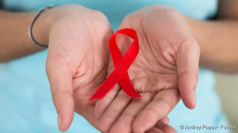
Leukemia (blood cancer): symptoms, causes, treatment
Leukemia (blood cancer): symptoms, causes, treatment
The term leukaemia refers to various cancers of the blood-forming system (“blood cancer”). What they all have in common is that degenerated white blood cells (leukocytes) multiply uncontrolled. The name already indicates this: “Leukemia” means “white blood”. Read more about the symptoms, causes, treatment and prognosis of leukemia here!
Leukaemia: short overview
- What is leukemia? Group of cancers of the haematopoietic system. Also called “blood cancer” or “leukosis”.
- Common forms: Acute myelogenous leukemia (AML), Acute lymphatic leukemia (ALL), Chronic myelogenous leukemia (CML), Chronic lymphatic leukemia (CLL; actually a form of lymph gland cancer)
- Possible symptoms: Fatigue and exhaustion, reduced performance, rapid fatigue, skin pallor, tendency to bleeding and bruising (haematomas), tendency to infections, fever of unknown origin, weight loss, night sweats, etc.
- Frequency: Every year, 13,700 people in Germany contract leukaemia, mostly between the ages of 60 and 70. Men are affected slightly more often than women. About four percent of the patients are children under the age of 15.
- Treatment options: depending on the type and stage of leukemia; e.g. chemotherapy, tyrosine kinase inhibitors, interferons, monoclonal antibodies, radiotherapy, stem cell transplantation, etc.
- Prognosis: Acute leukemia is often curable if it is detected and treated in time. In chronic leukemia, therapy can prolong the survival time of many patients. The only possible cure is a high-risk stem cell transplantation.
Leukemia: Symptoms
Leukemia can suddenly become apparent with symptoms and progress rapidly. Doctors then speak of acute leukemia. In other cases the blood cancer develops slowly and insidiously. Then it is a chronic leukemia.
How leukemia interferes with blood formation? In leukaemia the blood stem cells can no longer develop into functional blood cells.
Acute leukemia: symptoms
The symptoms of acute leukemia develop relatively quickly. Symptoms of both Acute Lymphocytic Leukemia (ALL) and Acute Myeloid Leukemia (AML) include
- reduced performance
- continued fever
- nocturnal sweating
- Fatigue
- Weight loss
- Bone and joint pain (especially in children with ALL)
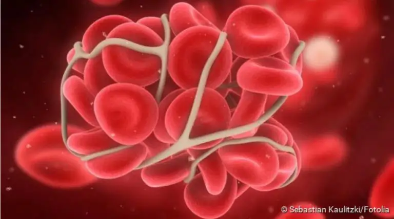
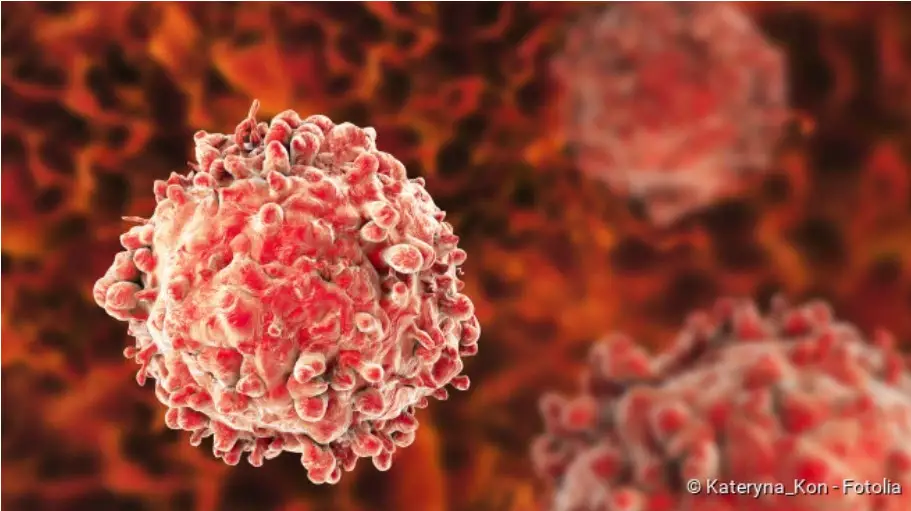
The patient’s body produces large quantities of immature white blood cells (leukocytes). These displace the healthy blood cells, i.e. mature leukocytes, red blood cells (erythrocytes) and blood platelets (thrombocytes). This causes more leukemia signs. For example, the lack of red blood cells leads to anaemia. The affected people suffer, for example:
The lack of blood platelets (thrombocytes) in acute leukemia often causes an increased tendency to bleed. For example, patients often suffer from bleeding gums or nose. In case of an injury it takes longer than usual for a wound to stop bleeding. In addition, patients increasingly get bruises (haematomas) – another typical sign. Severe platelet deficiency (thrombocytopenia) causes red bleeding into the skin, so-called petechiae.
Leukaemia can also weaken the immune system. As a result, patients often suffer from persistent infections such as poorly healing inflammations in the oral cavity. The reason: the patients’ bodies have too few functional white blood cells – and these are normally used to defend against infection. The immune system in leukaemia is therefore weakened overall.
Other possible leukemia symptoms are:
- lymph nodes swollen without pain
- enlarged liver and spleen
- Skin rashes
- Gum proliferation
Chronic leukemia: symptoms
Chronic leukemia begins insidiously. During the first months or even years many patients have no complaints at all. Some only report general symptoms such as fatigue and reduced performance. These are usually not recognized as signs of leukemia. This is why most patients do not go to the doctor. Only at an advanced stage do symptoms similar to an acute course develop in chronic leukemia.
In chronic myeloid leukaemia (CML) there are three phases in which the disease becomes increasingly aggressive. This can also be seen in the leukemia signs:
- Chronic phaseHere the number of white blood cells is abnormally high (leukocytosis) and the spleen is enlarged (splenomegaly). The latter can cause a feeling of pressure in the left upper abdomen. Other leukemia symptoms in this phase include fatigue and reduced performance.
- Acceleration phase (transition phase)The number of leukocytes is still rising. At the same time the number of red blood cells and platelets decreases. Typical symptoms of CML are now skin pallor, rapid heartbeat, shortness of breath and frequent nose and gum bleeding. Night sweats and fever can also occur. The liver is increasingly enlarged.
- Blast crisis (blast surge)In this last phase of the disease, the bone marrow releases large quantities of immature precursors of blood cells (so-called myeloblasts and promyelocytes) into the blood. This causes symptoms similar to those of acute leukemia. Usually the patients die soon.
Chronic lymphatic leukemia (CLL) also progresses only slowly. That is why the term “leukemia” is also in their name. Actually, however, it is not blood cancer but a special form of lymph gland cancer (malignant lymphoma).
Overview of the forms of leukaemia
The four main forms of leukemia are:
| Leukaemia form | Notes |
| Acute myeloid leukaemia (AML) | – begins quite suddenly and progresses rapidly
– most common acute leukemia – about half of the patients are older than 70 years |
| Chronic myeloid leukaemia (CML) | – slow, creeping progress (except in the last stage: blast crisis)
– mean age of onset of the disease at 50 to 60 years – in children very rare |
| Acute lymphatic leukemia (ALL) | – begins quite suddenly and progresses rapidly
– most common of all leukaemias – mainly in children (ALL is the most common type of cancer in children); adult patients mostly older than 80 years |
| Chronic Lymphocytic Leukemia (CLL) | – slow and creeping progress
– most frequent leukemia in adults; mean age of onset of the disease is between 70 and 75 years – does not belong to the “real” leukemias, but to lymph gland cancer (malignant lymphomas) |
There are also other types of leukemia, but these are very rare. One example is hairy cell leukemia.
Related to leukaemias are the so-called myelodysplastic syndromes (MDS). These are also chronic diseases of the bone marrow, in which too few functional blood cells are produced. The symptoms are similar to chronic myeloid leukemia. However, they are less pronounced in the beginning. In about 25 to 30 percent of patients, the myelodysplastic syndrome sooner or later develops into full-blown leukemia, namely acute myeloid leukemia.
Myelogenous leukaemia
Myeloid leukaemias originate from the so-called myeloid precursor cells in the bone marrow. These precursor cells normally develop into healthy red blood cells, platelets, granulocytes and monocytes. The last two are subgroups of white blood cells.
However, if myeloid precursor cells degenerate and start to grow uncontrolled, myeloid leukaemia develops. Depending on their course, physicians differentiate between Acute Myeloid Leukemia (AML) and Chronic Myeloid Leukemia (CML). Both forms of blood cancer mainly affect adults. AML is significantly more frequent than CML.
You can read more about the two forms of myeloid blood cancer in the article Myeloid leukemia.
Lymphatic leukemia
Lymphatic leukaemias originate from different blood cell precursors than myeloid blood cancer: this is where the so-called lymphatic precursor cells degenerate. Out of them against the lymphocytes. This subgroup of white blood cells is very important for the targeted (specific) defence against foreign substances and pathogens (specific immune defence).Allogeneic stem cell transplantation – for example with stem cells from family members – is a very effective but also risky therapy. It is therefore only used for certain forms of acute leukemia. And only if an unfavourable gene constellation suggests a poor prognosis. But then it can significantly improve the chances of recovery. One must weigh up with the patient whether the opportunities outweigh the risks.
Of course, a diagnosis like that kicks you out of life in the first place. But do not bury your head in the sand, try to find the best way for you. Don’t stay in bed, exercise, keep fit. A good network or self-help groups can also be helpful. You need a lot of support, get some.
Depending on the course of the disease, this is also referred to as acute lymphatic leukemia (ALL) or chronic lymphatic leukemia (CLM). ALL is the most common form of blood cancer in children and young people. In contrast, CLL typically occurs in older adults. It is only called “leukemia” (blood cancer) because of its course. Actually, CLL is a form of lymph gland cancer – it belongs to the so-called non-Hodgkin lymphomas.
You can read more about these two types of cancer in the article Lymphatic leukemia.
Hair-cell leukemia
Hair cell leukemia (or hair cell leukemia) is a very rare cancer. The same applies to it as to chronic lymphatic leukaemia: the part of the name “leukaemia” only indicates that the disease runs like blood cancer. However, it is attributed to lymph gland cancer (more precisely: non-Hodgkin lymphomas).
The part of the name “hair cells” comes from the fact that cancer cells have hair-like extensions.
Hair-cell leukemia only occurs in adulthood. Men suffer from it significantly more frequently than women. The chronic disease is not very aggressive. Most patients have a normal life expectancy.
You can read everything important about this cancer disease in the article Hair cell leukemia.
Leukemia in children
Leukemia is mainly a disease of adults: They account for about 96 percent of all patients. When leukaemia develops in children, it is almost always Acute Lymphatic Leukaemia (ALL). Acute myelogenous leukaemia (AML) comes in second place. Chronic leukemias are very rare in children.
If acute blood cancer is detected and treated early in childhood, the chances of recovery are good. In comparison, acute leukemias in adults have a rather poor prognosis.
Everything important about blood cancer in children can be found in the article Leukemia in children.
Leukemia: treatment
The leukemia treatment is individually adapted to each patient. Various factors play a role in this. In addition to the patient’s age and general state of health, this is mainly the course of the disease (acute or chronic).
Acute leukemia: treatment
Patients should start chemotherapy as soon as possible after the diagnosis of “acute leukemia”. It is considered the most important treatment method for acute blood cancer. The patient receives special medication, so-called cytostatics (chemotherapeutic drugs). They prevent cancer cells (and other rapidly dividing cells) from growing. The damaged cells can therefore no longer multiply. They are then recognised by the body’s own control mechanisms and are broken down in a targeted manner.
In most cases, the cytostatic drugs are administered directly into a vein (as an infusion), but occasionally they are also taken as tablets. They can be given individually or in combination and in different dosages. In this way, chemotherapy can be individually adapted to each patient. The treatment is also carried out in cycles: the patient receives the cytostatic drugs on one day or on several days in succession. This is followed by a treatment break (days to months) before a new cycle is started. Most cancer patients receive an average of four to six such chemotherapy cycles.
Basically, acute leukemia therapy consists of three phases, which together can last for months or years:
- Induction Therapy: Patients here receive a strong chemotherapy, which should eliminate as many cancer cells as possible and alleviate the most severe symptoms. The treatment is usually carried out as an inpatient in hospital.
- Consolidation therapy: It is designed to “consolidate” the success of induction therapy. To this end, many patients receive adapted chemotherapy to eliminate any remaining cancer cells.
- Maintenance therapy: The aim here is to stabilise the success of the treatment and prevent a relapse (recurrence). The maintenance therapy can be very different from patient to patient. Cytostatic drugs are often given in tablet form for at least one year.
Induction therapy can be so successful that practically no cancer cells can be detected in the patient’s blood and bone marrow. Doctors then speak of a remission. But it does not mean that the leukemia is cured. Individual cancer cells may still have survived. Therefore further therapeutic steps (consolidation therapy) are necessary.
The maintenance therapy is followed by the aftercare: The patient’s blood and bone marrow are examined regularly. If a relapse occurs, the cancer cells can be detected early in this way. In addition, follow-up care is also concerned with treating any side effects and long-term consequences of the previous chemotherapy.
Further therapy options
Sometimes a stem cell transplantation is also part of the leukemia treatment. Stem cells are the “mother cells” from which all blood cells in the bone marrow are created (and remain so for life). Prior to transplantation, high-dose chemotherapy (and possible whole-body radiation) destroys practically all of the patient’s bone marrow and (hopefully) all cancer cells. Afterwards, healthy stem cells are transferred to the patient as in a transfusion. The cells settle in the marrow cavities of the bones and produce new, healthy blood cells.
In leukaemia, stem cells are usually transferred from a healthy donor (allogeneic stem cell transplantation). More rarely, stem cells of the patient himself are concerned, which were taken from him before the bone marrow was destroyed (autologous stem cell transplantation). This method of therapy is particularly useful if other treatments (especially chemotherapy) are not effective enough or if the patient suffers a relapse.
Many patients with acute lymphatic leukemia (ALL) receive radiation therapy in addition to chemotherapy. On the one hand, the head is irradiated preventively, as cancer cells attack the brain more often. On the other hand, radiation can be used to treat malignant lymph nodes (for example in the breast area).
Chronic leukemia: treatment
Chronic Myeloid Leukemia (CML) is usually discovered in the chronically stable phase of the disease (see above). The doctor then usually prescribes so-called tyrosine kinase inhibitors (such as imatinib). These drugs act very specifically against blood cancer cells: They inhibit growth signals in the cells. This can stop the disease for many years. The tyrosine kinase inhibitors are taken as tablets, usually for life.
At the same time, the patients’ blood and bone marrow are regularly checked. If, for example, the patient’s blood values or condition deteriorate, this indicates that CML is moving on to the next phase (acceleration phase). The doctor then changes the drug treatment: he prescribes other tyrosine kinase inhibitors. In many patients, the disease can thus be returned to a chronically stable phase.
If this is not successful, an allogeneic stem cell transplantation might be considered – i.e. the transplantation of healthy, haematopoietic stem cells from a donor. So far, this is the only form of therapy that has a chance of completely curing chronic myeloid leukaemia. However, it is very risky. For this reason, the benefits and possible risks of treatment are carefully weighed for each patient beforehand.
At any stage of the disease, the condition of a patient can deteriorate significantly within a short period of time. Then doctors speak of a blast crisis. As in the case of acute leukemia, those affected usually receive intensive chemotherapy. In this way one tries to push back the signs of the disease as quickly as possible. Once the patient’s condition has improved and stabilized, a stem cell transplant may be useful.
Some patients with CML are treated with interferons. These are messenger substances with which the cells of the immune system communicate with each other. They can inhibit the growth of cancer cells. However, interferons – just like chemotherapy – are usually less effective in CML than the tyrosine kinase inhibitors described above.
However, this is not always true: Tyrosine kinase inhibitors work best in patients whose cancer cells have the so-called “Philadelphia chromosome”. This is the name given to a characteristically altered chromosome 22, which can be detected in more than 90 percent of all CML patients. The remaining patients do not have the altered chromosome. Therefore, treatment with tyrosine kinase inhibitors often does not work so well for them. Then it may be necessary to change the therapy and use interferons, for example.
Chronic Lymphocytic Leukemia (CLL) requires no treatment for many patients for a long time. Only when the blood values deteriorate or symptoms occur in an advanced stage do physicians initiate therapy – adapted to each individual patient.
For example, many patients receive chemotherapy plus so-called monoclonal antibodies (immunochemotherapy or chemoimmunotherapy): the artificially produced antibodies bind specifically to the cancer cells and thus mark them for the immune system. Both therapies are occasionally used separately.
If the cancer cells show certain genetic changes, treatment with tyrosine kinase inhibitors may be useful. These drugs block a pathologically altered enzyme that promotes the growth of cancer cells.
If other treatments do not work or there is a relapse later, doctors sometimes perform a stem cell transplant: Following aggressive chemotherapy, CLL patients are given healthy, haematopoietic stem cells from a donor (allogeneic stem cell transplantation). However, this risky treatment is only suitable for young or very fit patients.
Accompanying measures (supportive therapy)
In addition to leukemia treatment by means of chemotherapy, radiotherapy, etc., supportive measures are also very important. They serve, for example, to reduce symptoms of the disease and consequences of the treatment. This can enormously improve the well-being and quality of life of patients.
For example, nausea and vomiting are common and very unpleasant side effects of chemotherapy for leukemia (and other cancers). They can be relieved with special medication (antiemetics).
The increased susceptibility to infection is also a serious problem in leukemia. Both the disease itself and the chemotherapy weaken the immune system. It can then fight pathogens more poorly. This favours infections, which can then also be very difficult to treat. Sometimes they even become life-threatening! Therefore, careful hygiene and an environment with as few germs as possible are very important for leukemia patients. Many also receive antibiotics to prevent or treat bacterial infections. There are also special active ingredients, so-called antimycotics, for the treatment of fungal infections.
Other complaints can also often be treated specifically, for example anaemia by means of blood transfusion and pain with suitable painkillers.
Leukaemia: Causes and risk factors
The causes of the various forms of blood cancer have not yet been clearly clarified. However, experts have identified several risk factors that favour the development of leukaemia. These include:
Genetic predisposition: The risk of leukaemia is slightly increased if there has been a high incidence of cancer in the family. In addition, certain genetic diseases also make people more susceptible to blood cancer. For example, people with trisomy 21 (Down syndrome) have a 20-fold higher risk of developing acute myeloid leukaemia (AML) than people without this genetic change.
Age: The development of acute myeloid leukaemia (AML) is influenced by age: the risk of developing the disease increases with age. The same applies to chronic myeloid leukaemia (CML) and chronic lymphatic leukaemia (CLL). In contrast, acute lymphatic leukemia (ALL) occurs mainly in childhood.
Smoking: smoking is responsible for about ten percent of all leukemia cases, researchers estimate. For example, active smokers have a 40 percent higher risk of acute myelogenous leukaemia (AML) than people who have never smoked. Among former smokers, the risk of illness is still 25 percent higher.
Ionising radiation: These are various high-energy rays, for example radioactive rays. They damage the genetic material – especially in those body cells that divide frequently. This also includes the haematopoietic cells in the bone marrow. As a result, leukaemia can develop. The higher the radiation dose that affects the body, the greater the risk of leukaemia.
Such ionising radiation is also used in radiotherapy against cancer. Not only can they kill the cancer cells as desired, they can also damage the genetic material in healthy cells. In rare cases, patients develop a radiation-induced second cancer.
X-rays are also ionising. However, experts assume that an occasional X-ray examination cannot cause leukaemia. Nevertheless, x-rays should only be taken if absolutely necessary. This is because the damage that the rays cause in the body can accumulate in the course of life.
Chemical substances: Various chemical substances can increase the risk of leukaemia. These include benzene and other organic solvents. Pesticides (insecticides) and plant protection products (herbicides) are also suspected of promoting blood cancer.
This connection is certain for certain drugs that are actually used for the treatment of cancer (such as cytostatic drugs): they can promote the development of leukaemia in the long term. Before using such drugs, doctors therefore carefully weigh up the benefits and risks of such drugs.
Viruses: Certain viruses (HTL viruses I and II) are involved in the development of a very rare form of leukaemia. This so-called human T-cell leukemia mainly affects people in the Japanese region. This blood cancer variant is extremely rare in our country.
All other forms of leukaemia (AML, CML, ALL, CLL etc.) develop according to the current state of knowledge without any involvement of viruses or other pathogens.
Leukaemia: examinations and diagnosis
While chronic leukemia usually remains asymptomatic for a long time, acute forms start relatively suddenly and progress rapidly. However, symptoms such as reduced performance, paleness, palpitations, frequent nosebleeds or persistent fever also occur in many other and sometimes harmless diseases. Therefore they are not always taken seriously. With such complaints, however, there is always the suspicion of leukemia. That is why you should definitely see a doctor.
The first point of contact in case of suspected blood cancer is the family doctor. If necessary, he will refer the patient to a specialist, for example to a specialist in blood and cancer diseases (haematologist or oncologist).
Interview and physical examination
The doctor will first take the medical history (anamnesis). To do this, he asks how the patient feels in general, what complaints he has and how long they have existed. Information on any other diseases that currently exist or have occurred previously may also be important. In addition, the doctor will ask, for example, whether the patient is receiving any medication and whether cancer is known in the family.
This is followed by a thorough physical examination. Among other things, the doctor will listen to the lungs and heart, measure blood pressure and palpate the liver, spleen and lymph nodes. The results help the doctor to better assess the patient’s general condition.
Blood test
Blood tests are important if leukaemia or a related disease is suspected. A small blood count and a differential blood count are made. The small blood count indicates among other things the number of white blood cells (total number), red blood cells and platelets. For the differential blood count, the different subgroups of white blood cells are measured individually. In addition, the appearance of the blood cells can also be assessed under the microscope.
Pathologically altered blood values such as an increased number of white blood cells and a lack of red blood cells can be an important indication of leukemia. However, they can also have many other diseases as their cause.
In addition to the blood cells, other blood parameters are also assessed in the laboratory, for example kidney values and liver values. These values indicate how well these two organs work. If leukaemia is confirmed in the further course of the disease and the patient’s kidney and/or liver values are poor, this must be taken into account when planning therapy.
The laboratory also tests whether there are any signs of infection with bacteria, viruses or fungi in the blood. These germs could also be responsible for some complaints, such as an increased white blood cell count, fever and fatigue.
Bone marrow puncture
Whenever leukemia is suspected, it is necessary to examine the patient’s bone marrow in detail. For this purpose, the doctor uses a special needle under local anaesthetic to take a bone marrow sample, usually from the pelvic bone (bone marrow puncture). In the laboratory, the number and appearance of bone marrow cells are assessed. In typical changes, leukemia can be clearly identified. Sometimes even the form of the disease can be determined. In addition, the cells can be examined for changes in their genetic material (for example, the “Philadelphia chromosome” in chronic myeloid leukemia).
Adults and older children usually receive a local anaesthetic before the bone marrow is removed. For smaller children a short anaesthesia can be useful. The entire puncture usually takes only about 15 minutes and can be performed on an outpatient basis.
Further investigations
If the diagnosis of leukemia is confirmed, further examinations are often necessary. They should show whether other body regions and organs are also affected by the cancer cells. The general condition of the patient can also be better assessed with such examinations. This is important for therapy planning.
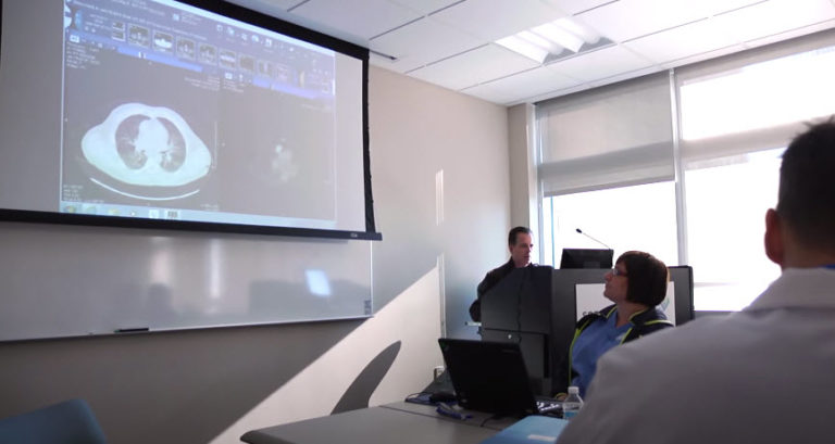
For example, internal organs (spleen, liver, etc.) can be examined using ultrasound. A computer tomography (CT) may also be performed. This imaging technique is also suitable for assessing the bones. This is important if the doctor suspects that the cancer cells have spread not only in the bone marrow but also in the bone itself. Sometimes a magnetic resonance tomography (MRT) or a scintigraphy is performed.
In acute lymphatic leukemia (ALL) and some subtypes of acute myeloid leukemia (AML), the cancer cells sometimes attack the brain or meninges. Possible signs of this are headaches as well as nervous failures such as impaired vision and paralysis. Then a sample of the spinal fluid can be taken (lumbar puncture) and analysed in the laboratory. An MRI can also be helpful in detecting cancer of the brain.
Leukaemia: course of disease and prognosis
For many people with leukaemia, the chances of survival are much better today than they were several years or decades ago. Modern therapies can often improve the chances of recovery. However, if the cancer is already too advanced, treatment can at least alleviate the symptoms of many patients and prolong their survival time.
In individual cases, the prognosis for leukaemia depends on various factors. First and foremost, the type of cancer and the stage of the disease at the time of diagnosis. The prognosis is also influenced by how well the patient responds to the therapy. Other factors that influence life expectancy and chances of recovery from leukaemia are the age and general condition of the patient and any concomitant diseases.
Leukemia: chances of recovery
“Is leukemia curable?” Many patients and their relatives ask themselves this question. In principle, there is a cure for acute leukemia. The earlier the disease is discovered and treated, the greater the chances of recovery. This is especially true for younger patients:
Without treatment, most patients survive the diagnosis of acute leukemia for only about three months. With treatment, 95 percent of children and 70 percent of adults with acute lymphatic leukemia (ALL) are still alive five years after diagnosis. In acute myeloid leukemia (AML), the 5-year survival rate is 40 to 50 percent in patients under 60 years of age and 20 percent in the 60+ age group.
Even if the cancer can be pushed back, a relapse (recurrence) may occur later, even after months and years. Especially in the case of an early relapse the chances of recovery decrease. Leukemia patients must then be treated again. Sometimes doctors choose a more aggressive therapy or other treatment methods.
In chronic leukemia, cancer cells multiply more slowly than in acute forms of cancer (exception: blast crisis in CML) – and usually for years. Therefore, the treatment is usually less intensive, but must be continued in the long term. In general, chronic leukaemia cannot be cured (the only chance of a cure is a risky stem cell transplant). In many patients, however, the therapy can alleviate the symptoms and slow down the progression of chronic leukemia.