COPD: symptoms, causes, consequences, therapy
COPD: Brief overview
- Main cause: smoking (chronic smoker’s cough)
- Typical symptoms: Shortness of breath, coughing, sputum
- Complications: frequent infections of the bronchial tubes and pneumonia, cor pulmonale, emphysema
- Important research: Lung function test, blood gas analysis, X-ray examination of the chest (X-ray thorax)
- Treatment options: Depending on the COPD stage stop smoking, medication, sports, respiratory and physiotherapy, long-term oxygen therapy, operations (incl. lung transplantation)
COPD: Description
COPD is often played down as “smoker’s lung” or “smoker’s cough”. However, COPD is a serious lung disease, which once started, progresses and often leads to premature death.
COPD is widespread: In Germany, about ten percent, or eight million people, suffer from this. Experts predict that this number will rise to ten million by 2020. COPD is one of the most frequent causes of death.
COPD: Definition and important terms
What exactly is COPD? The abbreviation stands for the English term “chronic obstructive pulmonary disease”. In technical terminology “chronic obstructive bronchitis” (COB). The terms “chronic bronchitis” and “emphysema” are associated with COPD:
Chronic bronchitis: According to the WHO, chronic bronchitis is present when coughing and sputum (productive cough) exist for at least three months in two consecutive years. One speaks of a “simple chronic bronchitis” if coughing and sputum occur only once due to overproduction of mucus in the lungs. At this stage, the changes in the lungs can still recede if the cause (for example, smoking) is eliminated. If this does not happen, chronic bronchitis can develop into COPD.
COPD: Lung disease is usually a combination of chronic obstructive bronchitis and emphysema – hence the term “chronic obstructive pulmonary disease”. The changes in the lungs cannot be completely reversed at this stage.
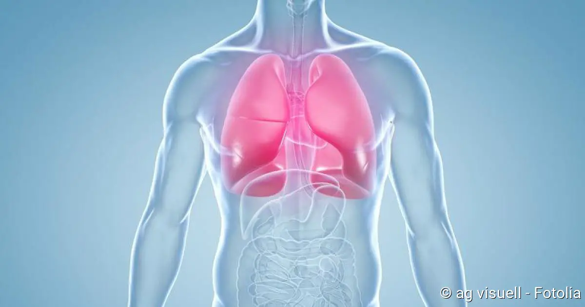
Pulmonary emphysema: Pulmonary emphysema is an over-inflated lung. In the course of COPD, the wall structure of the alveoli (alveolar septums) can be destroyed, which irrevocably expands the air space. The lung then no longer looks like a vine with grapes (as in a healthy person), but like a large balloon. Physicians speak of pulmonary emphysema (over-inflation of the lungs).
Exacerbated COPD: The term exacerbation stands for a sudden, intermittent worsening of COPD. Symptoms such as chronic coughing, shortness of breath and mucous sputum increase acutely. Exacerbations can be a stressful and threatening event for patients. Exacerbated COPD is a sign that lung function is deteriorating rapidly. Infection-exacerbated COPD is additionally accompanied by a viral or bacterial infection.
COPD: causes and risk factors
COPD can have various causes. In the vast majority of cases it is “home-made” – by smoking.
Main Cause: Smoking
The main cause of COPD is active or passive smoking. About 90 percent of all COPD patients are smokers or ex-smokers. Lungs and bronchial tubes suffer most from the constant nicotine consumption. The risk of COPD in smokers and ex-smokers is seven times higher compared to people who have never smoked. About 20 percent of long-term cigarette smokers develop COPD. Every second smoker over 40 years of age suffers from “smoker’s cough”. Men are significantly more frequently affected than women.
Alpha-1-antitrypsin deficiency
Smoking is not always the cause of the complaints. In some patients, chronic lung disease is due to a genetic deficiency of the protein alpha-1-antitrypsin (AAT): This blood protein inactivates so-called proteases – enzymes that are supposed to break down destroyed tissue during inflammatory processes. However, in people with AAT deficiency, the proteases can damage lung tissue unchecked. The result is a chronic inflammation with narrowing of the bronchial tubes, just like in COPD triggered by pollutants. As the disease progresses, pulmonary emphysema may develop. In addition, AAT deficiency can cause liver damage and promote cirrhosis of the liver.
AAT deficiency is quite common in the United States, and about as common in Europe as type 1 diabetes, but does not receive the same attention. Therefore, the hereditary disease is often not recognized and treated in time.
A COPD is not acutely dangerous. However, if ignored over many years, it can become threatening due to the progressive disruption of breathing. In this respect, early detection (lung function test by a doctor) and effective measures in early stages are of particular importance.
For people with COPD, it is true that every day of smoking worsens lung function and thus breathing a little bit. The moment you stop, the remaining lung function is maintained and may even improve slightly. If smoking continues, however, the shortness of breath will steadily worsen.
The most effective advice is to stop smoking, the earlier the better! The second most important is regular exercise. It is just as important to avoid any infections that could worsen this disease in the long term. As a holistic physician I did not only rely on medication for treatment, but also on nutrients and a healthy intestine. A lot of vegetables and fruit and few dairy products are crucial.
Other causes of COPD
Another possible cause of COPD is air pollution. Nitrogases and sulphur dioxide (SO2) play a particularly important role here. Studies have shown that living on busy roads with high levels of particulate matter increases the risk of COPD. Frequent infections during childhood also increase the probability of developing COPD.
The causes of chronic lung disease can also be harmful dusts, vapours, smoke or gases to which some people are exposed at work. In non-smokers, the risk of developing COPD later on is 2.4 times higher. The risk of illness is even 18 times higher for smokers.
A very rare cause of COPD is the congenital lack of antibodies (antibody deficiency syndrome).
COPD: Mechanism of origin
The starting point of COPD is usually an obstructive bronchitis: Inhaled pollutants inflame the small airways, the bronchioles. The lungs secrete increased amounts of mucus for protection. The mucus is normally transported by the cilia towards the exit (pharynx). These are very fine, flexible hairs (cilia) on the surface of special cells that line most of the respiratory tract (ciliated epithelium).
However, pollutants such as nicotine destroy the cilia, so that the ciliated epithelium gradually loses its ability to clean and transport. Finally, it is replaced by a more resistant squamous epithelium, which causes the lung tissue to thicken. The wall of the alveoli thus becomes thin and unstable on exhalation. If the affected person tries to exhale with all his strength, the alveoli collapse. Ultimately, this process leads to a permanent narrowing of the airways. The consequences are shortness of breath and poor performance.
COPD: a vicious circle
People who are unable or unwilling to stop smoking encourage the vicious circle of COPD. Basically it is determined by two factors:
On the one hand, the excessively produced mucus can block the small airways. As a result, the negative pressure of the inhaled air barely reaches the alveoli. When breathing out, however, the air can no longer be forced out completely so that residual air remains in the bubbles. With the next breath more residual air remains in the bubbles. Thus the pressure in the lungs slowly increases. As a result, the small alveoli of the lungs connect with each other and thus become larger and larger expanded bubbles, so-called emphysema bubbles. Overall, this reduces the surface area of the bubbles.
On the other hand, inhaled pollutants and lung infections destroy the lung tissue. Proteases and protease inhibitors play a role in this. Proteases are enzymes that break down proteins and can therefore be cell-damaging. Protease inhibitors in turn inhibit the cleavage of protein and thus have a protective effect. Alpha-1-antitrypsin is the most important protease inhibitor. Lung infections lead to the release of more proteases. Nicotine in turn disables the protease inhibitors so that they cannot perform their protective function. Due to this imbalance of proteases and protease inhibitors, the lung tissue becomes more unstable and the wall structure of the small alveoli (alveolar septa) perishes.
Consequences of COPD
COPD has serious consequences:
For the lungs, COPD means a loss of elasticity and an increase in residual volume (volume of air that remains in the lungs after exhalation and cannot be exhaled arbitrarily).
The small airways are increasingly obstructed and place extreme demands on the respiratory muscles. This increases the amount of carbon dioxide in the blood (hypercapnia).
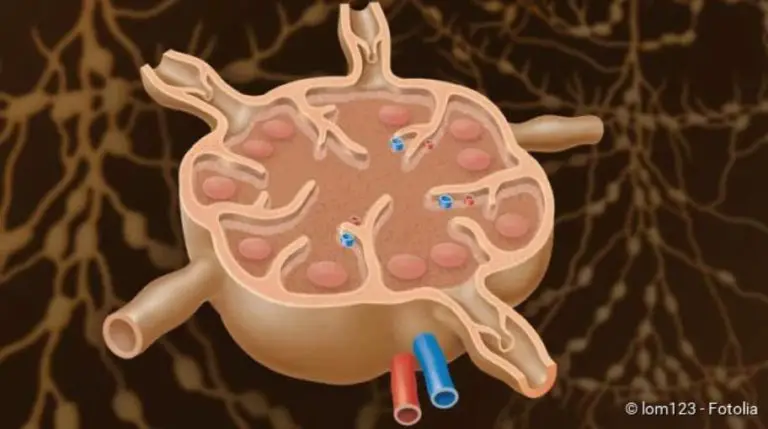
With the alveolar septa, the pulmonary capillaries are also lost. These are tiny connecting vessels between arteries and veins. As the airways are insufficiently ventilated, less oxygen reaches the blood vessels. As a result, the blood vessels constrict and the lungs are ultimately less supplied with blood. This mechanism is also called the Euler-Liljestrand reflex by physicians. It prevents blood from passing through the lungs without being supplied with oxygen. Due to the narrowed pulmonary arteries, the blood pressure in the pulmonary circulation increases, which in turn puts a strain on the right heart. The consequence is a so-called cor pulmonale (lung heart). It causes breathing difficulties under stress or already at rest.
COPD: Symptoms
On average, the lungs of an adult person hold five to six litres. Without physical exertion we breathe in and out only half a liter of it. The lungs therefore have relatively large reserves. A creeping deterioration of their function therefore usually goes unnoticed for years. Initial COPD symptoms are often neglected by those affected as a “smoker’s cough” that they cannot get rid of. However, it is important for the course of the disease to recognise and treat COPD symptoms early on.
By the way: If COPD is caused by an AAT deficiency and the lungs are additionally stressed by smoking or infections, the disease progresses faster than normal. Typical symptoms of COPD then appear much earlier.
Typical COPD symptoms
Typical COPD symptoms are coughing and sputum. Respiratory distress also occurs, initially only on exertion, especially when the COPD symptoms worsen acutely (exacerbation). Blue lips or fingers are signs of cyanosis, i.e. a reduced oxygen supply due to deteriorating lung capacity.
The most important COPD symptoms are summarized here:
- Respiratory distress, initially only under stress, later also at rest.
- A cough that gets worse and more persistent over time.
- Sputum, which is getting tougher and tougher and more difficult to cough up.
Following the characteristic COPD symptoms of breathlessness-cough-ejection, many doctors speak of AHA symptoms.
How does the smoker’s cough develop?
Chronic coughing (smoker’s cough) in smokers is one of the characteristic symptoms of COPD and a risk factor for deterioration in lung function. It is created as follows:
The cilia in the lungs are gradually destroyed by smoking and lose their cleaning function. Therefore the body has to cough up the secretion that has accumulated, especially at night. In the morning after getting up, the typical smoker’s cough is therefore particularly tormenting. In the course of the day, many patients are then relatively free of complaints. The sputum has a greyish colour in smokers.
Symptoms of COPD: Complications
COPD is a chronic, progressive disease. You should therefore see your doctor regularly. If you are largely symptom-free, it is sufficient to have an examination once a year. However, if your condition worsens (increase in coughing, sputum and/or shortness of breath), you should consult your doctor immediately. In this way, deterioration and complications can be detected and treated in time:
Infections and shortness of breath
Long-term COPD typically results in recurrent bronchial infections and pneumonia. The reduced lung function can also lead to constant shortness of breath.
Cor pulmonale
In the late stage of COPD, the so-called cor pulmonale can occur: The right half of the heart enlarges and loses functional strength – a right heart weakness develops. The consequences of this are, among other things, accumulation of water in the legs (edema) and abdomen (ascites) and congested neck veins. Heart failure and failure of the respiratory muscles are severe, life-threatening complications.
Drum flail finger and watch glass nails
On the hands, so-called drum flail fingers with watch glass nails can occur. These are rounded finger endings with arched fingernails. They are the result of a reduced oxygen supply.
Pulmonary emphysema and fascia
If COPD progresses, lung tissue is destroyed and an over-inflated lung (emphysema) develops. This is often expressed by a grasping thorax. The chest is shaped like a barrel and the front ribs are almost horizontal. The grasping thorax is one of the typical symptoms of emphysema.
COPD symptoms: Pink puffer and Blue Bloater
According to the external appearance of COPD sufferers, two types can be distinguished in principle: the “Pink Puffer” and the “Blue Bloater”. These are two clinical extremes; in reality, mainly mixed forms occur:
| Type | Appearance |
| Pink puffer | With the “pink puffer” the pulmonary emphysema is in the foreground. The over-inflated lung leads to constant shortness of breath, which overstrains the muscles that assist breathing. The affected person therefore consumes an extremely large amount of energy. The typical “pink puffer” is therefore underweight. A dry cough occurs occasionally. The oxygen levels in the blood are not reduced because enough carbon dioxide is breathed out. The most common cause of death is respiratory failure. |
| Blue bloater | The “blue cough” (also called “bronchitis type”) suffers mainly from coughing and sputum, COPD is the main symptom. He is usually overweight and cyanotic, i.e. his lips and nails have a bluish discoloration due to lack of oxygen. Nevertheless, the shortness of breath is only slightly pronounced. The “Blue Bloater” has an increased risk of right heart failure. |
COPD symptoms of exacerbation
In the course of COPD, there can always be an acute worsening of the COPD symptoms – called exacerbation. Exacerbations can be divided into three degrees of severity: light, moderate and severe. The symptoms of COPD go beyond the normal level of daily fluctuation and usually last longer than 24 hours.
Responsible for an acute worsening of COPD symptoms are, for example, viral and bacterial infections, air pollution (smog), damp-cold weather, accidents with injuries to the ribcage, as well as drugs that have a negative effect on breathing.
Are signs of worsening COPD symptoms
- Respiratory distress increase
- Increase in coughing
- Sputum increase
- Change in colour of the sputum (yellow-green sputum is a sign of a bacterial infection)
- general malaise with fatigue and possibly fever
- Breast Strait
Are signs of severe exacerbation:
- Respiratory distress at rest
- reduced oxygen saturation in the lungs (central cyanosis)
- Use of the respiratory auxiliary musculature
- Water retention in the legs (edema)
- Loss of consciousness to coma
The symptoms of COPD are more frequent in autumn and winter. Every acute exacerbation means a potential danger to life for the affected person, because with increasing oxygen deficiency and exhaustion of the respiratory muscles, the lungs can fail within a short time! Those affected with acute worsening of COPD symptoms should therefore urgently seek medical attention – they need more intensive treatment.
COPD stages
At the end of 2011, a new classification of COPD was presented by the GOLD (Global initiative for chronic Obstructive Lung Disease). Previously, only the restriction of lung function and symptoms were decisive for the GOLD-COPD stages.
The GOLD classification from 2011 onwards also took into account the frequency of a sudden worsening of COPD (exacerbation rate) and the outcome of patient questionnaires when classifying stages.
Finally, in 2017, GOLD has revised its recommendations again. Although the same parameters are still taken into account, the COPD stages are now classified more precisely.
COPD stages: Classification until 2011
There are four stages of COPD in total. The classification is based on lung function, which is measured with the spirometer. The one-second capacity (FEV1) is determined. This is the largest possible lung volume that can be exhaled within one second.
| Severity |
Symptoms | One second capacity (FEV1) |
| COPD 0 Risk group |
chronic symptoms :cough, sputum |
inconspicuous |
| COPD 1 lightly |
with or without chronic symptoms :coughing, sputum, shortness of breath during heavy physical exertion |
unobtrusive (not below 80 percent of the setpoint) |
| COPD 2 medium |
with or without chronic symptoms :coughing, sputum, shortness of breath |
limited (between 50 and 80 percent of the setpoint) |
| COPD 3 heavy |
with or without chronic symptoms :coughing, sputum, shortness of breath |
limited (between 30 and 50 percent of the setpoint) |
| COPD 4 extremely heavy |
chronically insufficient oxygen supply | severely restricted (below 30 percent of the setpoint) |
COPD 1
A one-second capacity below 80 percent of the normal value is called mild COPD, i.e. COPD grade I. Typical symptoms are usually chronic coughing with increased mucus production. However, it is also possible that both symptoms are absent. Breathlessness is usually not yet noticed. Often the affected people do not even know that they have COPD.
COPD 2
COPD grade II is a moderate COPD. Breathing difficulties may occur under heavy physical strain. The symptoms are usually more pronounced, but can also be absent completely. The one-second capacity is between 50 and 80 % of normal value. Patients who are not physically active may not even notice the worsening of the disease.
COPD 3
This stage of COPD is already a severe COPD and many alveoli are already no longer functional. The one-second capacity is between 30 and 50 percent of normal value for patients with COPD grade III. The symptoms of coughing and sputum are increasingly noticeable, and even slight exertion can make people lose their breath. But there are also patients who do not have a cough or sputum.
COPD 4
If the one-second capacity is below 30 percent of the normal value, the disease is already well advanced. The patient is in the final stage of COPD, i.e. COPD grade IV. The oxygen level in the blood is very low, which is why patients suffer from respiratory distress even at rest. As a sign of end-stage COPD, right heart damage may already have developed (cor pulmonale).
COPD stages: Classification from 2011
The classification of COPD-GOLD stadiums from 2011 onwards continued to be based on lung function, measured by one-second capacity. In addition, the GOLD now also took into account the frequency of exacerbations and symptoms recorded by means of a questionnaire (COPD assessment test) such as shortness of breath or limited physical fitness. According to the new findings, four patient groups were identified: A, B, C and D.
The measured one-second capacity initially roughly determined whether a patient was assigned to groups A/B (with COPD 1 or 2) or C/D (with COPD 3 or 4). The severity of the symptoms as well as the number of exacerbations finally determined whether it was stage A or B or C or D.
Example: A patient with a one-second capacity of between 50 and 80 percent of normal value would therefore correspond to a COPD stage GOLD 2 and would therefore be allocated to Group A or B. If he had severe symptoms of COPD, he would be assigned to group B, with only mild symptoms in group A. Similarly, the classification for groups C and D is made for one-second capacities below 50 percent (GOLD 3 and 4).
COPD stages: COPD assessment test
The COPD assessment test (CAT) is a questionnaire that helps you and your doctor to assess the impact of COPD on your quality of life. The test takes only a few minutes and consists of eight questions, for example, whether you cough, have phlegm or are restricted in your domestic activities. The extent to which the individual points apply is indicated by points between zero and five. Overall, a total score between 0 and 40 can be achieved. For the stage classification of a COPD it depends on whether the patient scores more or less than ten points.
Determination of COPD stages since 2017
Since 2017, GOLD has been dividing the COPD stadiums even more finely. In contrast to the 2011 classification, the one-second capacity (still classified as GOLD stages 1 to 4) is now shown separately and in addition to groups A to D. This allows a more precise classification and thus a better adapted treatment.
Example: While according to the 2011 classification, a patient with a one-second capacity of less than 50 percent automatically already belonged to groups C or D, this is not necessarily the case according to the new classification. If he has only minor complaints and at most one exacerbation per year, he may even belong to group A. However, the one-second capacity still plays an important role and is given additionally. With a one-second capacity of 40 percent of normal value (Gold 3), the example patient would correspond to a COPD level GOLD 3A according to the new classification.
As has been the case since 2011, the doctor determines the severity of the symptoms with the help of the CAT questionnaire.
The new COPD stages are designed to ensure that each patient receives the optimal treatment for his or her individual needs.
| Staging according to FEV1 | & | Group allocation according to complaints and exacerbations | ||
| 1
2 3 4 |
A | B | ≤1 Exaz./ year | |
| C | D | ≥ 2 Exac. / year | ||
| CAT < 10 | CAT ≥ 10 | |||
FEV1 = One-second capacity in lung function testCAT
= COPD assessment test (result in the symptom questionnaire)
COPD: examinations and diagnosis
If COPD is suspected, your family doctor will usually refer you to a lung specialist (pneumologist) first. Whether you actually suffer from the lung disease COPD or another disease can be found out with special examinations. Especially the distinction between COPD and asthma is very important because the symptoms are very similar.
COPD diagnosis: First examinations
The doctor will first ask you about your medical history (anamnesis). It can give first indications of an existing COPD. Possible questions from the doctor are:
- How long have you been coughing and how often?
- Do you cough up more phlegm, maybe especially in the morning? What colour is the mucus?
- Do you experience shortness of breath under stress such as climbing stairs? Has this one already appeared in peace?
- Do you smoke or have you been smoking? If so, how long and how many cigarettes a day?
- What do you do for a living? Are you exposed to pollutants in the workplace?
- Has your efficiency decreased?
- Have you lost weight?
- Do you suffer from other diseases?
- Do you have symptoms such as water retention (edema) on your legs?
This is followed by a physical examination: If there is a COPD, the doctor will hear certain breathing noises such as wheezing during exhalation when listening to the lungs with a stethoscope. Often, a weakened breathing sound can be heard, which is also called “silent lung” by doctors. This occurs when the lungs are over-inflated (pulmonary emphysema) because the patient is then no longer able to breathe out the respiratory volume. If the lungs are slimy, damp rales can be heard. When tapping the lung, a hollow sounding (hypersonic) knocking sound occurs when the lung is over-inflated.
In addition, the doctor looks for signs of reduced oxygen supply (for example, blue lips or fingers = cyanosis) and heart failure (for example, water retention in the ankle area).
How do COPD and asthma differ?
COPD and asthma are not easy to distinguish. Asthma is a chronic inflammatory disease of the airways caused by hypersensitivity or allergy. A certain trigger then leads to a narrowing of the airways, which manifests itself as shortness of breath. The narrowed airways can recede spontaneously or through treatment. Asthma usually manifests itself in childhood or early adulthood.
In COPD, on the other hand, the disease develops insidiously; it is also not an allergy. In contrast to asthma, this narrowing of the airways can only be partially, but not completely, improved by medication.
COPD Diagnosis: Apparative examinations
For a COPD test, various examination methods are used. Lung function tests (LuFu for short) such as spirometry, whole body plethysmography and blood gas analysis are performed to see how well the lungs are working. Pulmonary function tests are mainly used to diagnose COPD and to assess the course and therapy of the disease.
In spirometry, the patient breathes through the mouthpiece of the spirometer, which measures the respiratory volume. The vital capacity and one-second air are measured, which are parameters for lung function. The one-second capacity (FEV1) is the largest possible lung volume that can be forcibly exhaled within one second. The vital capacity (FVC) is the total lung volume that can be exhaled after deep inhalation. If the one-second capacity is less than 70 per cent of the normal value, it is considered to be COPD.
In whole-body body plethysmography, the patient sits in a closed cabin and breathes through a tube of the spirometer. Respiratory resistance and lung capacity are determined. Whole-body body plethysmography can be used to distinguish COPD from other diseases such as asthma.
An analysis of the blood gases shows the oxygen content in the blood. Especially in patients under 45 years of age with additional emphysema, a targeted search is made for alpha-1-antitrypsin deficiency. Experts recommend that this examination be performed once in a lifetime for every COPD patient. The determination can be performed from a single drop of blood – similar to a blood glucose measurement. If it results in a congenital deficiency, the diagnosis can be supplemented by a genetic test to detect the genetic change (mutation) underlying the AAT deficiency.
Some lung and heart diseases cause similar symptoms to COPD. To confirm the diagnosis, an X-ray examination, a computer tomography (CT) and an ECG can be performed. X-ray and CT can be used to detect such things as pneumonia, congestion of the lungs, pneumothorax and tumours. The ECG provides information about the heart function. There may be indications of increased pulmonary pressure (pulmonary hypertension) and thus right-heart strain.
COPD: Therapy
The COPD therapy is a long-term therapy. It depends on the severity of the disease. Overall, COPD therapy comprises drug and non-drug measures and has the following objectives:
- Increase of physical resilience
- Symptom relief
- Prevention of acute deterioration (exacerbations)
- Improvement of the state of health and quality of life of the person concerned
- Avoidance of complications
COPD therapy: stop smoking
Most COPD patients are smokers. The most important component of COPD treatment is the renunciation of nicotine.
The stop smoking should be approached with medical and psychosocial support. You can give yourself additional motivation is by considering the effects of COPD related to smoking:
According to a scientific study, in COPD patients the stop smoking stabilizes lung function compared to those who continue to smoke. In the first year, lung function even increased again in former smokers. Coughing and sputum improved. Young smokers in particular benefited from the smoking ban. Patients who had stopped smoking also had a lower mortality rate.
However, these positive changes only occur if nicotine is completely abstained from. It is not enough for effective COPD therapy to simply smoke less than before.
COPD Therapy: Training
As part of COPD therapy, patients are advised to attend COPD training whenever possible. There, they learn everything about the disease, its self-control as well as the correct inhalation technique and breathing, for example, breathing with pursed lips (lip brake) In the COPD training, patients also learn how to recognize and treat an acute worsening (exacerbation) in time. It has even been proven that patient education in people with mild and moderate COPD improves the quality of life and reduces the number of exacerbations and thus the number of hospital stays per year. Such training courses are therefore important elements of COPD therapy and are offered by many health insurance companies.
COPD therapy: Drugs
Various groups of active ingredients are used as COPD drugs. They can alleviate the symptoms and delay the progression of the disease through various mechanisms.
COPD therapy: bronchodilators
Bronchodilators are bronchodilating drugs that are very often used in COPD therapy. They reduce shortness of breath on exertion, reduce the number of exacerbations, help against inflammation and allow the mucous membrane to subside.
Physicians distinguish between short-acting and long-acting bronchodilators. Long-acting bronchodilators are superior to short-acting ones in COPD therapy, more effective and easier to use. They only need to be taken once or twice a day and are therefore well suited for regular use.
Bronchodilators include anticholinergics, beta-2 sympathomimetics and theophylline.
Anticholinergics: The best known representative is the short-acting ipratropium. It dilates the bronchial tubes, reduces the production of mucus, improves breathing and thus physical performance. The full effect occurs after 20 to 30 minutes.
The effect of the long-acting anticholinergic tiotropium bromide lasts 24 hours. The active ingredient is therefore only taken once a day. It reduces lung hyperinflation, shortness of breath, exacerbations and hospitalization. Other long-acting anticholinergics are aclidinium bromide and glycopyrronium bromide.
Beta-2 sympathomimetics: In acute respiratory distress, short-acting beta-2 sympathomimetics are used. They work almost immediately. The substances used are called fenoterol, salbutamol and terbutaline.
Long-acting beta-2 sympathomimetics such as salmeterol and formoterol have a duration of action of about twelve hours, indacaterol even about 24 hours. The active ingredients help against shortness of breath both during the day and at night. In addition, they improve lung function, reduce hyperinflation of the lungs and decrease the number of exacerbations. This improves the quality of life of COPD patients. Cardiac arrhythmia can occur as side effects.
Theophylline: This active substance dilates the bronchial tubes in the long term. It is only used in the treatment of COPD when a common drug combination such as anticholinergics plus beta-2 sympathomimetics is not sufficient. The problem with taking the drug is that the drug level can fluctuate, which greatly increases the risk of side effects. Doctors must therefore frequently check the theophylline level in the blood (blood level). Because of the risks mentioned, theophylline is controversial and rather a reserve drug. It should only be used as a third choice in COPD therapy.
Combinations of bronchodilators: If the above-mentioned active ingredients do not work sufficiently when used individually, inhalable slow-acting bronchodilators (such as tiotropium) and beta-2 sympathomimetics can be combined. The bronchodilator effect is thereby enhanced. This can also be useful if, for example, the beta-2 sympathomimetic drug has too strong side effects such as racing heart and trembling. Its dose can be reduced by combining it with an anticholinergic. This reduces the risk of side effects.
COPD therapy: Cortisone
In addition to bronchodilators, cortisone (for inhalation) is one of the frequently used active ingredients in COPD therapy. In long-term therapy, it prevents the inflammatory tendency of the respiratory tract and can thus prevent acute exacerbations. Cortisone is used especially for patients who suffer from asthma in addition to COPD.
The use of inhaled cortisone will be considered where the one-second capacity is less than 50 per cent of normal value and where additional steroids and/or antibiotics are used for exacerbations. The risk of side effects is low with this form of application.
Cortisone in tablet form is not recommended for long-term COPD therapy.
COPD Therapy: Mucolytics
Expectorants/mucolytics are not generally recommended for COPD therapy. They are only used in cases of massive mucus and acute infections. Regular inhalation with salt solutions is also helpful in this case. Bacterial infections usually require additional treatment with antibiotics.
COPD patients should also make sure to drink enough – but not too much! This can put additional strain on the lungs and promote the derailment of a chronic cor pulmonale.
COPD therapy: Inhalation systems
Various inhalation systems are available for COPD therapy. Besides metered dose inhalers and powder inhalers, nebulisers are also used.
Inhaling a drug has the advantage that the active substance can easily reach the diseased parts of the lungs. As a result, the patient can breathe more easily because the active ingredients relax the smooth muscles in the walls of the bronchial tubes and thus reduce the muscle tone in the bronchi. The lungs are then less over-inflated. Typical COPD symptoms such as shortness of breath, coughing and sputum are relieved.
COPD therapy depending on the stage of the disease
The guideline of the German Respiratory League recommends that COPD therapy should be a step-by-step adapted treatment, depending on the stage of the disease. From stage to stage, more COPD drugs must be used in addition.
At first, one starts with short-acting bronchodilators, which are only used when needed. If the symptoms increase, long-term bronchodialators complement the therapy. Only when COPD progresses further and the symptoms increase significantly, do doctors additionally prescribe inhaled cortisone. In principle, however, one tries to avoid cortisone preparations as long as possible. In the final stage of the disease, long-term oxygen therapy is usually necessary. A surgical intervention (emphysema surgery) can also be considered.
COPD therapy: Vaccinations
Since people with COPD often suffer from infections, vaccinations against influenza and pneumococcus are recommended – regardless of the severity of the disease. Vaccination has been shown to reduce mortality and should therefore always be considered.
COPD Therapy: Exacerbations
Depending on the severity of the exacerbation and the patient’s impairment, COPD therapy is carried out on an outpatient or inpatient basis.
In some cases it is sufficient to increase the drug dose. If the symptoms such as coughing, shortness of breath and sputum still increase, you must talk to your doctor. Other warning signs are fever and yellow-green sputum. They are indications of an infection that can be treated with antibiotics. If there is no improvement, an inpatient COPD therapy is necessary.
Patients with severe exacerbation (severe shortness of breath, FEV1 < 30 percent, rapid deterioration, advanced age) must generally seek inpatient treatment in a hospital.
COPD Therapy: Rehabilitation
COPD patients experience increasing shortness of breath under stress. That’s why most people move less and less. The consequences: The muscles deteriorate, the ability to bear weight is reduced, and those affected become increasingly inactive and ultimately immobile. In addition, the quality of life decreases and social contacts are avoided. This can lead to depression and, as a consequence, further deterioration in breathing.
Targeted physical training is therefore very important in COPD. It can prevent the degradation of musculature and resilience. For COPD therapy, there are various rehabilitation programmes such as lung sports or respiratory and physiotherapy.
COPD Therapy: Sport
Physical training increases the patient’s quality of life and resilience. In addition, the number of exacerbations is decreasing. Physical training such as endurance and weight training should therefore be an integral part of long-term COPD therapy. Training programs of four to ten weeks, in which the patients complete three to five exercise units per week under supervision, show particularly positive effects. In a lung sports group, for example, you can learn specific exercises that strengthen the respiratory muscles and thus make breathing easier.
COPD therapy: respiratory and physiotherapy
Here, COPD patients learn how to relieve difficult breathing at rest and under stress with special breathing techniques and certain postures. You will learn how to make the chest more flexible and how to cough up fixed mucus more easily. This improves the ventilation of the lungs. At the same time, optimal respiration prevents infections of the respiratory tract, from which COPD patients often suffer. The slack abdominal muscles are also systematically trained. The correct breathing behaviour is important because it takes away the feeling of anxiety in case of shortness of breath, increases self-confidence and improves performance.
Well-known body positions that make breathing easier are the so-called coachman’s seat and the lip brake.
Carriage seat: Support yourself with your arms on your thighs or on a table top so that the entire chest can support the exhalation. Close your eyes and breathe calmly and evenly. The coach seat reduces increased airway resistance and supports the function of the respiratory muscles. In this position, the weight of the shoulder girdle is also taken off the chest.
Lip Brake: Breathe out as slowly as possible against the pressure of your loosely closed lips. This causes the cheeks to puff up a little. With this technique the respiratory flow is slowed down and the bronchi remain open. The lip brake increases the pressure in the lungs and thus prevents the airways from collapsing during exhalation.
COPD therapy: Long-term oxygen therapy
In the advanced stage of COPD, the oxygen supply through the damaged lung is no longer sufficient. The patient therefore suffers from permanent shortness of breath. Then long-term oxygen therapy is useful; the patient receives oxygen bottles from which he inhales oxygen via a nasal probe. In this way, the oxygen concentration in the blood is stabilized, and breathing difficulties are reduced. When long-term oxygen therapy is applied for 16 to 24 hours a day, the prognosis improves in patients with chronic shortness of breath.
COPD therapy: diet and weight
Weigh yourself regularly to check that your weight remains stable. Many COPD patients show unwanted weight loss. This can be a sign of an unfavourable course of the disease. Sometimes a targeted nutritional therapy is then necessary in order to put on a few kilos again. If breathing difficulties are the reason for eating too little, smaller and more frequent meals are advisable.
On the other hand, sudden weight gain can also occur. It is usually an indication of heart failure (more precisely: right heart failure). The cardiac output is then no longer sufficient to ensure normal blood circulation. The blood accumulates, causing water to pass from the vessels into the tissue, where it is stored (edema). This can happen at the ankles, for example. Affected patients should limit their calorie intake to 1200 to 1500 kilocalories per day in order to lose weight successfully.
COPD therapy: aids
In advanced COPD, many patients can no longer cope with their everyday life without help. In some cases, autonomy can be maintained by means of aids. These include, for example, extensions for shoehorn and brushes and mobile walking aids (walkers).
COPD Therapy: Surgery
For patients with advanced COPD, who are increasingly suffering from pulmonary hyperinflation and for whom neither medication nor rehabilitation measures can help, surgery is considered. There are different surgical methods that can be used in COPD therapy:
Bullectomy
In a bulllectomy, functionless pulmonary alveoli are removed. The balloon-like dilated bronchi (bullae) no longer participate in the gas exchange and push away neighbouring healthy lung tissue. If the bullae take up more than a third of a lung wing, their removal can improve lung function and relieve respiratory distress.
Before a bullectomy, a bronchoscopy, a series of pulmonary function tests and a computer tomography of the lungs are performed.
Lung volume reduction
In lung volume reduction, so-called lung valves are inserted endoscopically into the airways of the over-inflated lung sections. These valves close when inhaled and open when exhaled. In this way, air can flow into the over-inflated areas and old air can escape again. This is intended to reduce pulmonary hyperinflation, relieve respiratory distress and improve lung function.
However, pulmonary volume reduction is only possible for a special form of emphysema (heterogeneous form). Preliminary examinations are used to determine whether or not this form of COPD therapy is suitable in individual cases.
Lung Transplantation
COPD is the most common reason for lung transplantation. On average, 60 COPD patients undergo lung transplantation each year. This surgical measure of COPD therapy can prolong life and improve the quality of life.
A lung transplantation comes into question when all other COPD therapy measures (long-term oxygen therapy, home respiration, etc.) have been exhausted and the life expectancy of the patient is clearly limited according to the experts’ assessment. On average, patients have to wait about two years for a new lung.
Admission criteria for getting on the waiting list for a lung transplant are, for example
- abstinence from smoking for at least six months
- One-second capacity below 25 % of normal value
- Pulmonary hypertension (pulmonary hypertension)
- respiratory global insufficiency (disturbed gas exchange in the lungs, which reduces the oxygen partial pressure in the blood and increases the carbon dioxide partial pressure in the blood)
Exclusion criteria are considered to be too high a risk of complications in lung transplantation. One such exists:
- severe overweight (BMI over 30 kg/m²)
- Coronary heart disease (CHD)
- Renal failure
- Cirrhosis of the liver
- Age over 60 years (in exceptional cases: 65 years)
COPD: Course of the disease and prognosis
The prognosis of COPD depends on whether the progression of the lung disease can be slowed down. The most important element is to refrain from smoking. This has a positive effect on the symptoms, the course of the disease and life expectancy in COPD patients.
Non-obstructive bronchitis is often still curable if smoking is avoided or exposure to harmful substances is avoided. Already a few hours after the last cigarette the smoker’s lungs improve. The regeneration of the lungs is already visible from two weeks on: the blood circulation has improved and the lung capacity has increased.
With COPD lungs, however, it is usually too late and lost lung tissue cannot be recovered. In such cases, however, an effective drug therapy can significantly reduce the symptoms. If a lung transplantation is successful, COPD is curable. However, the affected person must then take drugs that suppress the body’s own immune system for the rest of their life. Otherwise the new lung will be rejected.
COPD: Prevention
To prevent the development of COPD, you should first and foremost stop smoking. About 90 percent of all COPD patients have smoked for a long time or are still doing so.
In addition, the following tips apply:
- Both in your leisure time and at work, make sure that you are not exposed to harmful influences such as dusty, cold or polluted air more than necessary. This also includes the avoidance of rooms polluted by tobacco smoke.
- Get vaccinated against flu (influenza) and pneumococcus.
If you already have COPD, the following measures will help to prevent COPD exacerbation (acute worsening)
- If you still smoke, give it up. This significantly reduces the risk of exacerbations.
- Take part in a patient training course. There you will learn how to deal with COPD and how to adjust your medication dose in case of an acute worsening. In this way, the number of hospital stays can be reduced.
- Exercise regularly.
- Get vaccinated against flu (influenza) and pneumococcus.
- Do breathing exercises (like a coachman’s seat). It improves the breathing technique, the ventilation of the lungs and thus the oxygen supply. At the same time, optimal respiration prevents infections of the respiratory tract from which COPD patients suffer.
- Have your back patted (tapping massage). This promotes the expectoration of mucus in smokers’ lungs.
- Do not stay in smoky rooms. Avoid places heavily contaminated with pollutants (dust, smoke).
- Contact the company physician if there is a high level of pollution in the workplace. Seek medical attention immediately!
- Pay attention to your diet and weight. Every excess kilo is a burden on the body. Conversely, underweighting also worsens the forecast. A healthy diet also supports the immune system.
- Support your immune system also by avoiding harmful factors such as stress.
- Drink enough water and inhale regularly with salt water. It helps to cough up the phlegm.
With these measures you can positively influence the course of COPD and your quality of life.
COPD: Life expectancy
Life expectancy in COPD patients depends, among other things, on how much the airways are narrowed. In general, the stronger the narrowing, the worse the prognosis.
In addition, there are a number of factors that influence the life expectancy of patients. For example, nicotine consumption, age and possible concomitant diseases play a role.
You can read everything important about the life expectancy of COPD patients in the article COPD Life Expectancy.
Further information
Book recommendations:
- Breathing deeply at last: Effective breathing exercises for asthma, bronchitis and COPD (Rainer Dierkesmann, 2015, TRIAS)
- Nutrition in COPD (Agnes Budnowski, 2015)